by
John R. Fischer, Senior Reporter | October 14, 2019
The other proposal calls for combining SPECT with CT to help determine treatment plans for lung cancer. Patients with this disease often experience trouble breathing which can be made worse by the delivery of radiotherapy to the lungs, and lead to side effects such as disabling breathlessness. This makes it extremely important for doctors to limit the amount of normal lung tissue regions exposed to radiation.
SPECT, which produces computerized 3D scans by tracking and imaging the location of a radioactive tracer compound, can show certain body functions such as blood flow to tissues and organs. Scanning 48 patients with it, researchers were able to predict if a person’s lung function would deteriorate following radiotherapy based on how much lung tissue received radiation, even at low dose. Those who became breathless were found to have a larger volume of healthy lung tissue exposed to low dose radiation, while those who did not report increased trouble breathing were found with SPECT to have comparatively small regions of healthy lung exposed to radiation.


Ad Statistics
Times Displayed: 365084
Times Visited: 6976 Quality remanufactured Certified Centrifuges at Great prices! Fully warranted and backed by a company you can trust! Call or click for a free quote today! www.Centrifugestore.com 800-457-7576
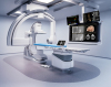
SPECT and CT can help determine treatment
for lung cancer patients whose breathing is
limited already prior to receiving radiotherapy
The researchers say that together, SPECT scans and standard CT may provide helpful insights for treatment of patients whose breathing is limited already before receiving radiotherapy, and help amend treatment plans to limit low dose radiation exposure to normal regions of lungs with good blood flow.
“The data from our study confirm a significant difference in dose–volume histograms between anatomic (defined on CT) and perfused lung (defined on perfusion SPECT) volumes,” said the authors in their study. “This is partly explained by technical differences in the acquisition of lung volume between the techniques, but shows the potential for using perfusion volumes as an independent “organ-at-risk” when determining radiotherapy dose scheduling.”
The study on cervical cancer was published in the
International Journal of Radiation Oncology, Biology, Physics.
The study on lung cancer was published in
The British Journal of Radiology.
Back to HCB News