
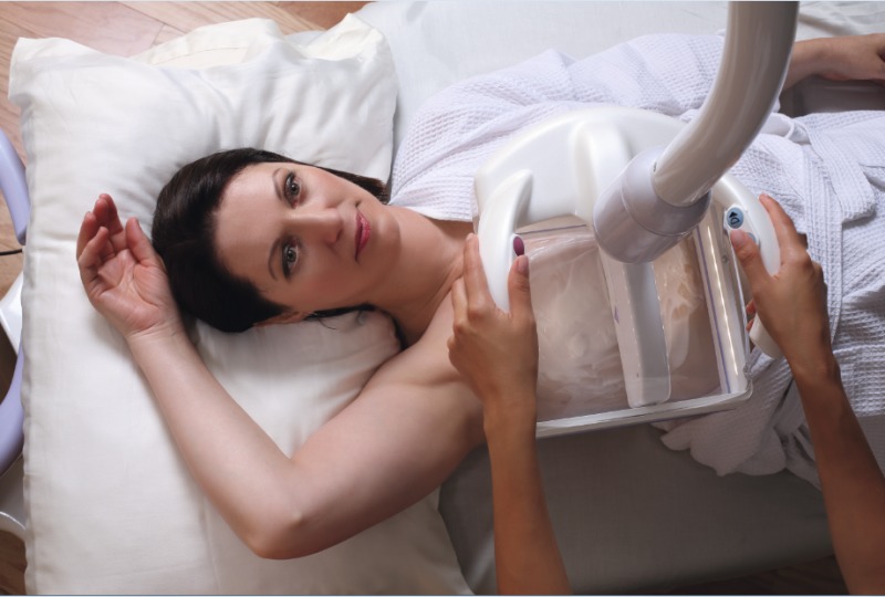
Siemens' ACUSON S2000 ABVS
Dense breast legislation and technological advances drive automated breast ultrasound
July 10, 2017
by Lauren Dubinsky, Senior Reporter
Nancy Cappello followed all the normal guidelines to monitor for, and protect against, developing breast cancer.
From eating right and exercising, to performing self exams and having annual mammography scans, she was as diligent as you could reasonably expect someone to be.
So, when she was diagnosed with stage 3 breast cancer in 2004, and discovered the disease had been growing undetected due to the shortcomings of mammography for imaging patients like her who have dense breast tissue, she decided to do something about it.
Cappello advocated in her home state of Connecticut for a law that required health care providers to educate women on their breast density as well as potential options for supplemental screening. In 2009, it became the first state in the U.S. to require providers to offer supplemental whole-breast ultrasound to women with dense breasts and to mandate that insurers cover the additional screening.
A number of states followed suit. To date, a total of 31 states require some level of breast density notification following a mammogram.
Since dense breast tissue can hide cancer on mammograms, supplemental screening options such as ultrasound or tomosynthesis are vital for this patient population. A 2012 study published in JAMA found that ultrasound detects an average of four more cancers per 1,000 women screened compared to mammography.
A 2014 study published in Diagnostic Imaging: Breast revealed that more than 85 percent of cancer diagnoses only detected by screening ultrasound are invasive or node negative.
The Adjunct Screening With Tomosynthesis or Ultrasound in Women With Mammography-Negative Dense Breasts prospective trial from 2016 was the first to compare ultrasound and tomosynthesis as adjunct screening options. It found that the added cancer detection yield of 7.1 per 1,000 screens for ultrasound was significantly higher than that from tomosynthesis at 4 per 1,000 screens.
MR can also be used as a supplemental screening tool in the dense breast population. A study involving average-risk women indicated that MR has a much higher cancer detection rate than ultrasound or tomosynthesis — more than 22 per 1,000 in year one and 7.5 per 1,000 in subsequent years.
“Breast MR is a very powerful tool,” says Sankar Suryanarayanan, general manager of the ABUS business at GE Healthcare. “It has extremely high sensitivity, but the limitations are that it involves a gadolinium contrast injection, which has been associated with toxicity. Some women have a problem going into the MR scanner because they are claustrophobic and [it’s expensive].”
He adds that breast MR is used as an adjunct screening tool when the woman’s overall risk is 20 percent or more, based on family history and BRCA gene tests. Alternatively, the only risk factor that’s needed to use ultrasound is dense breast tissue.
Handheld vs. automated ultrasound
When it comes to breast ultrasound, there are two options — conventional handheld breast ultrasound or automated breast ultrasound (ABUS). The main difference between the technologies is the skill that’s required of the operator.
SonoCiné’s Automated Whole Breast Ultrasound (AWBUS), which received FDA clearance in 2008, has the patient in a supine position as an articulating robotic arm moves a handheld ultrasound over the breast. This system combines a conventional high-resolution 2-D handheld transducer with an automated scanning arm.
Siemens Healthineers and U-Systems took a different approach and developed standalone ABUS systems that use high-frequency transducers.
U-Systems’ somo-V, which was approved by the FDA for screening purposes in 2012, is equipped with a larger transducer that’s similar in size and shape to a mammography compression paddle. The operator positions the transducer over the breast and presses a button to begin the acquisition.
Siemens’ ACUSON S2000 Automated Breast Volume Scanner was introduced to the market in 2009. A typical exam consists of three automated one-minute scans of each breast in an anterior-posterior position and both breasts in an oblique position.
“Traditional handheld ultrasound is a great technology, but there are some limitations,” says Srish Sinha, head of the women’s health segment at Siemens. “The transducer is a small footprint, so when you are scanning the breast you have to take small pieces of images and reconstruct them in your head, so it’s very subjective.”
He adds that handheld ultrasound is also highly operator dependent. If the operator is a good sonographer, then good data is obtained, but if not, then the quality suffers.
GE’s Suryanarayanan notes that some facilities may not be able to afford the human resources to scan patients every day with handheld ultrasound technology. He believes that ABUS can fill that need because the operator doesn’t have to be as skilled.
“The machine does it for them very similarly to mammography,” he says. “It makes the process repeatable, efficient and it delivers the same level of quality across patients, day in and day out, regardless of the type of operator.”

The Invenia ABUS
GE Healthcare acquired U-Systems in November 2012 and developed its Invenia ABUS, which got the FDA nod in June 2014. It’s currently the only ABUS system that can be used as a screening adjunct to mammography.
The European Asymptomatic Screening Study, which was published in the European Journal of Radiology in October 2016, found that the Invenia ABUS increases breast cancer detection by 57 percent. The analysis involved 1,668 women between the ages of 40 and 74 who underwent 15-minute ABUS exams after digital mammography screening.
The findings were equivalent to 6.6 cancers detected per 1,000 women compared with the 4.2 cancers detected using mammography alone. In addition, 23 women who underwent mammography alone were recalled for further testing, but only 15 women who received the supplemental ABUS exam were recalled.
Dr. Jason Davis, radiologist and director of imaging at OSF Medical Group in Illinois, has been using the Invenia ABUS as a supplemental screening tool for women with dense breasts since February 2016. He prefers it over conventional handheld ultrasound.
“Handheld ultrasound used for screening was shown to be good, but the problem is that it consumes a lot of time and resources, and screening exams are not supposed to be that way,” he says.
He believes ABUS gives the patient and reading radiologist another level of confidence. He explains that mammography detects only 50 percent of breast cancer in women with dense breasts, but adding ultrasound can bring that rate up to 90 percent.
“When [dense breast] cancers are finally detected, they tend to be larger cancers at a higher stage,” he says. “We would like to catch those at the size they are when cancers are found with mammography.”
There isn’t a dedicated screening reimbursement code for ABUS like there is for mammography, but facilities can use the code for bilateral breast ultrasound to receive reimbursement
More than 200 facilities worldwide have purchased the Invenia ABUS and over 120 are in use in the U.S.
The state of the market
iVu Imaging Corporation, a small start-up company based in Texas, developed an ABUS system called SOFIA that’s different from other systems in that the patient is placed in the prone position. The company designed it that way in order to capture the entire breast at one time, instead of acquiring multiple views of the breast from different angles.
During the exam, the patient’s breast is coated with a warm acoustic lotion and placed into a funnel with a built-in transducer. SOFIA then takes 52 seconds to scan the entire breast and the entire exam, including acquisition and interpretation, takes about 10 minutes.
In December 2014, iVu announced that Hitachi Aloka Medical America, Inc. (HAMA) would be the exclusive distributor of SOFIA in North America. As part of the agreement, iVu used HAMA’s wide market reach to launch SOFIA to the masses.
Philips Healthcare also became involved in the ABUS market after signing an agreement with SonoCiné in February 2014 to incorporate the company’s AWBUS technology into its EPIQ and iU22 ultrasound systems.
The global ABUS market is flourishing. A Global Market Insights report from January estimated that the market will exceed $2 billion by 2024. Factors driving the market include the rising prevalence of breast cancer and programs and government initiatives that are creating greater awareness of breast density.
Certain segments of the market are expected to witness substantial growth through 2024. China’s market share made up over 60 percent of regional revenue in 2015 and is anticipated to reach approximately $1 billion by 2024.
Brazil is expected to grow at an annualized rate of 20 percent during the forecast period and surpass $60 million by 2024.
“The space is very attractive now for adjunct breast screening and we are seeing a lot of new entrance in this space,” says GE’s Suryanarayanan. “GE still continues to be the largest player, but we have seen a lot of smaller players coming in with their own versions of ABUS.”
Mammography remains the gold standard for breast cancer screening, but Suryanarayanan says researchers and many of GE’s customers are investigating whether ABUS could be a primary screening tool for women with dense breasts.
However, a large-scale clinical trial that demonstrates the performance of each modality on a population level will be required for guidelines to change.
“From what we can see, it seems to make sense because where mammography is weak, ABUS is strong,” says Suryanarayanan. “This whole notion of personalized breast care is the path in which the industry is moving.”
From eating right and exercising, to performing self exams and having annual mammography scans, she was as diligent as you could reasonably expect someone to be.
So, when she was diagnosed with stage 3 breast cancer in 2004, and discovered the disease had been growing undetected due to the shortcomings of mammography for imaging patients like her who have dense breast tissue, she decided to do something about it.
Cappello advocated in her home state of Connecticut for a law that required health care providers to educate women on their breast density as well as potential options for supplemental screening. In 2009, it became the first state in the U.S. to require providers to offer supplemental whole-breast ultrasound to women with dense breasts and to mandate that insurers cover the additional screening.
A number of states followed suit. To date, a total of 31 states require some level of breast density notification following a mammogram.
Since dense breast tissue can hide cancer on mammograms, supplemental screening options such as ultrasound or tomosynthesis are vital for this patient population. A 2012 study published in JAMA found that ultrasound detects an average of four more cancers per 1,000 women screened compared to mammography.
A 2014 study published in Diagnostic Imaging: Breast revealed that more than 85 percent of cancer diagnoses only detected by screening ultrasound are invasive or node negative.
The Adjunct Screening With Tomosynthesis or Ultrasound in Women With Mammography-Negative Dense Breasts prospective trial from 2016 was the first to compare ultrasound and tomosynthesis as adjunct screening options. It found that the added cancer detection yield of 7.1 per 1,000 screens for ultrasound was significantly higher than that from tomosynthesis at 4 per 1,000 screens.
MR can also be used as a supplemental screening tool in the dense breast population. A study involving average-risk women indicated that MR has a much higher cancer detection rate than ultrasound or tomosynthesis — more than 22 per 1,000 in year one and 7.5 per 1,000 in subsequent years.
“Breast MR is a very powerful tool,” says Sankar Suryanarayanan, general manager of the ABUS business at GE Healthcare. “It has extremely high sensitivity, but the limitations are that it involves a gadolinium contrast injection, which has been associated with toxicity. Some women have a problem going into the MR scanner because they are claustrophobic and [it’s expensive].”
He adds that breast MR is used as an adjunct screening tool when the woman’s overall risk is 20 percent or more, based on family history and BRCA gene tests. Alternatively, the only risk factor that’s needed to use ultrasound is dense breast tissue.
Handheld vs. automated ultrasound
When it comes to breast ultrasound, there are two options — conventional handheld breast ultrasound or automated breast ultrasound (ABUS). The main difference between the technologies is the skill that’s required of the operator.
SonoCiné’s Automated Whole Breast Ultrasound (AWBUS), which received FDA clearance in 2008, has the patient in a supine position as an articulating robotic arm moves a handheld ultrasound over the breast. This system combines a conventional high-resolution 2-D handheld transducer with an automated scanning arm.
Siemens Healthineers and U-Systems took a different approach and developed standalone ABUS systems that use high-frequency transducers.
U-Systems’ somo-V, which was approved by the FDA for screening purposes in 2012, is equipped with a larger transducer that’s similar in size and shape to a mammography compression paddle. The operator positions the transducer over the breast and presses a button to begin the acquisition.
Siemens’ ACUSON S2000 Automated Breast Volume Scanner was introduced to the market in 2009. A typical exam consists of three automated one-minute scans of each breast in an anterior-posterior position and both breasts in an oblique position.
“Traditional handheld ultrasound is a great technology, but there are some limitations,” says Srish Sinha, head of the women’s health segment at Siemens. “The transducer is a small footprint, so when you are scanning the breast you have to take small pieces of images and reconstruct them in your head, so it’s very subjective.”
He adds that handheld ultrasound is also highly operator dependent. If the operator is a good sonographer, then good data is obtained, but if not, then the quality suffers.
GE’s Suryanarayanan notes that some facilities may not be able to afford the human resources to scan patients every day with handheld ultrasound technology. He believes that ABUS can fill that need because the operator doesn’t have to be as skilled.
“The machine does it for them very similarly to mammography,” he says. “It makes the process repeatable, efficient and it delivers the same level of quality across patients, day in and day out, regardless of the type of operator.”
GE's Invenia ABUS
The Invenia ABUS
GE Healthcare acquired U-Systems in November 2012 and developed its Invenia ABUS, which got the FDA nod in June 2014. It’s currently the only ABUS system that can be used as a screening adjunct to mammography.
The European Asymptomatic Screening Study, which was published in the European Journal of Radiology in October 2016, found that the Invenia ABUS increases breast cancer detection by 57 percent. The analysis involved 1,668 women between the ages of 40 and 74 who underwent 15-minute ABUS exams after digital mammography screening.
The findings were equivalent to 6.6 cancers detected per 1,000 women compared with the 4.2 cancers detected using mammography alone. In addition, 23 women who underwent mammography alone were recalled for further testing, but only 15 women who received the supplemental ABUS exam were recalled.
Dr. Jason Davis, radiologist and director of imaging at OSF Medical Group in Illinois, has been using the Invenia ABUS as a supplemental screening tool for women with dense breasts since February 2016. He prefers it over conventional handheld ultrasound.
“Handheld ultrasound used for screening was shown to be good, but the problem is that it consumes a lot of time and resources, and screening exams are not supposed to be that way,” he says.
He believes ABUS gives the patient and reading radiologist another level of confidence. He explains that mammography detects only 50 percent of breast cancer in women with dense breasts, but adding ultrasound can bring that rate up to 90 percent.
“When [dense breast] cancers are finally detected, they tend to be larger cancers at a higher stage,” he says. “We would like to catch those at the size they are when cancers are found with mammography.”
There isn’t a dedicated screening reimbursement code for ABUS like there is for mammography, but facilities can use the code for bilateral breast ultrasound to receive reimbursement
More than 200 facilities worldwide have purchased the Invenia ABUS and over 120 are in use in the U.S.
The state of the market
iVu Imaging Corporation, a small start-up company based in Texas, developed an ABUS system called SOFIA that’s different from other systems in that the patient is placed in the prone position. The company designed it that way in order to capture the entire breast at one time, instead of acquiring multiple views of the breast from different angles.
During the exam, the patient’s breast is coated with a warm acoustic lotion and placed into a funnel with a built-in transducer. SOFIA then takes 52 seconds to scan the entire breast and the entire exam, including acquisition and interpretation, takes about 10 minutes.
In December 2014, iVu announced that Hitachi Aloka Medical America, Inc. (HAMA) would be the exclusive distributor of SOFIA in North America. As part of the agreement, iVu used HAMA’s wide market reach to launch SOFIA to the masses.
Philips Healthcare also became involved in the ABUS market after signing an agreement with SonoCiné in February 2014 to incorporate the company’s AWBUS technology into its EPIQ and iU22 ultrasound systems.
The global ABUS market is flourishing. A Global Market Insights report from January estimated that the market will exceed $2 billion by 2024. Factors driving the market include the rising prevalence of breast cancer and programs and government initiatives that are creating greater awareness of breast density.
Certain segments of the market are expected to witness substantial growth through 2024. China’s market share made up over 60 percent of regional revenue in 2015 and is anticipated to reach approximately $1 billion by 2024.
Brazil is expected to grow at an annualized rate of 20 percent during the forecast period and surpass $60 million by 2024.
“The space is very attractive now for adjunct breast screening and we are seeing a lot of new entrance in this space,” says GE’s Suryanarayanan. “GE still continues to be the largest player, but we have seen a lot of smaller players coming in with their own versions of ABUS.”
Mammography remains the gold standard for breast cancer screening, but Suryanarayanan says researchers and many of GE’s customers are investigating whether ABUS could be a primary screening tool for women with dense breasts.
However, a large-scale clinical trial that demonstrates the performance of each modality on a population level will be required for guidelines to change.
“From what we can see, it seems to make sense because where mammography is weak, ABUS is strong,” says Suryanarayanan. “This whole notion of personalized breast care is the path in which the industry is moving.”