
Siemens Healthineers' SOMATOM go.Up.
Rethinking CT in the value-based era
September 11, 2017
by Lauren Dubinsky, Senior Reporter
When spectral CT was first introduced roughly a decade ago, adoption was slow. However, it’s recently been gaining steam and even landed a spot in ECRI Institute’s 2016 Top 10 Hospital C-Suite Watch List.
"The real advantage of spectral CT is that it gives you an extra dimension of information so you can start looking at what chemicals and elements are within the anatomy,” says Jason Launders, director of operations in the Health Device Group at ECRI.
One downside of this technology is its high price tag. According to ECRI, a spectral CT scanner costs between $1.6 million and $2.2 million, and that doesn’t include the ongoing maintenance expenses.
In addition, several challenges stand in the way of widespread adoption. Reimbursement is not provided for spectral CT and the technology's workflow is not efficient.
In order for radiologists to evaluate these new types of images, they have to do extra work. They only have a few minutes to read each study, and if it’s going to take longer than that, then they’re not going to do it.
These scanners are costly because they are equipped with advanced dose-reduction features and the latest image reconstruction and display tools. They also have a fast gantry rotation and are capable of large volumetric imaging.
"I think we are in the early days with this [technology]," says Launders. "Manufacturers are beginning to put tools in the hands of radiologists so they can do more of this image manipulation in a way that suits their workflow. The key word here is convenience."
A new generation of spectral CT systems
In August 2016, Siemens Healthineers scored FDA clearance for its SOMATOM Drive spectral CT system. While the previous generation, SOMATOM Force, had a price tag making it suitable primarily for academic centers, the Drive was designed for facilities with smaller budgets.
“The Drive takes the technical and clinical benefits of the Force and brings them into a platform that is more accessible from a cost of ownership standpoint,” says Matthew Dedman, CT marketing director for Siemens.
It can be used for a wide range of clinical applications including pediatrics, cardiology, oncology and emergency medicine. It’s also equipped with the company’s Straton MX Sigma X-ray tubes and Sigma generators, which allow for more targeted beam focusing.
GE Healthcare announced in February that it received FDA clearance for its GSI Xtream technology for its Revolution CT. According to the company, it’s the first volume spectral CT technology designed to improve small lesion detection, tissue characterization and metal artifact reduction with a simplified workflow.
GSI Xtream is between two and eight times faster than GE’s previous generation GSI technology.
“The benefits of spectral imaging have been widely published, but it has not been broadly adopted because of the workflow limitations in the industry,” says Scott Schubert, general manager of premium CT at GE. “We think GSI Xtream is now the first technology that is able to make spectral imaging a routine application.”
Philips Healthcare’s flagship spectral CT system, the IQon Spectral CT, received FDA clearance in November 2014. It’s different from the GE and Siemens systems in that the clinicians don’t have to decide up-front whether they want spectral results for the patient.
“Spectral results are available for every patient scanned on the IQon, which helps to increase diagnostic confidence, as well as provide the opportunity to be a cost-effective resource by reducing the need for downstream testing,” says Lakshmi Gudapakkam, general manager of the diagnostic imaging business group at Philips.
The system uses a dual-layered detector, which is essentially two detectors sandwiched on top of each other. The advantage of that technique is that a spectral imaging is generated every time, according to Launders.
He adds that it's still the early adoption days of spectral CT and that its true potential remains unknown.

The cost-effective side of premium CTs
At last year’s Radiological Society of North America annual meeting, Toshiba Medical introduced its new Aquilion ONE / GENESIS Edition premium CT. The system filters the X-ray beam in a way that reduces dose and improves image quality.
Its PUREViSION Optics, PUREViSION Detector and FIRST reconstruction technology improve high contrast spacial resolution up to 129 percent while reducing dose by about 85 percent compared to the Aquilion ONE ViSION Edition.
Reconstruction times have been improved by up to 80 images per second and FIRST reconstruction is achieved in as little as three minutes per 320 images.
Three systems have been installed to date – the first at the National Institutes of Health and the other two at St. Cloud Hospital in Minnesota and Cincinnati Children’s Hospital Medical Center.
Tim Nicholson, senior manager of CT market development at Toshiba Medical, says that customers are willing to purchase premium CT systems if they can leverage them to improve patient outcomes and reduce costs.
“We hear of value-based medicine and we know radiology wants to show value,” he adds. “Customers are asking for something that proves you can use that technology and bring value to the patient's care and the institution that’s using it.”
In May, Toshiba Medical and the Stroke Care Center (SCC) at the Gates Vascular Institute in Buffalo, N.Y., published the results of their multi-year study on the economic impact of using the previous generation Aquilion ONE CT to diagnose and treat acute stroke.
The researchers gathered inpatient baseline data before the CT was installed between July and September 2007. Once the CT was installed, the 2009 to 2012 data revealed a significant increase in using CT-based protocols for stroke diagnoses, which translated into a reduction in health care costs and better patient outcomes.
Within the first five years after the Aquilion ONE was installed, SCC raked in more than $5.4 million in cost savings. That largely was due to reduced length of stays, less outpatient services and complications and lower readmission rates.
Toshiba Medical believes that any health care facility with a dedicated stroke program can reap the same cost savings.
More affordable options
"Health providers looking to enhance their CT capabilities may be faced with challenges such as reimbursement cuts, patient populations with low ability to pay or inefficiencies related to growth and expansion," says Philips' Gudapakkam.
In response to these trends, Philips introduced its Access CT system in March. The system was specifically designed for health care organizations that want to establish or improve their CT imaging capabilities at an accessible cost.
The iDose technology helps to reduce the need for tube replacement, which is the highest recurring cost associated with CT ownership. It's a reconstruction algorithm that reduces exposure time and extends tube life.
The iFlow console workflow platform offers features that simplify and automate the technologists’ routine, which helps them achieve consistently high image quality. It also helps the providers maintain and increase their referral base.
"[Access CT] expanded our capabilities into emerging markets and for cost-conscious health care organizations," says Gudapakkam. "As health care needs increase across the globe, many health care organizations are increasingly challenged to efficiently diagnose and treat a greater number of patients."
In April, Siemens received FDA clearance for its lower cost of ownership SOMATOM go. CT platform, which includes the SOMATOM go.Now and SOMATOM go.Up CT systems. It was designed as a single-room concept so facilities can save on installation costs and it has a smaller footprint compared to the company’s other CT platforms.
It’s primarily for routine CT work including head, chest, abdomen and pelvic exams, which make up 50 to 60 percent of CT exams. It’s ideal for community hospitals, critical access hospitals as well as outpatient imaging centers.
Siemens reported that the first few installations were underway in July and the company predicted that by the end of August approximately five would be installed in the U.S.
Personalized imaging for RT planning
The invention of CT played a crucial role in the development of radiation therapy planning. Other modalities like MR and PET are used secondarily in the treatment planning process, but CT remains the only 3-D imaging modality used for dose calculation, according to an article published in the journal BioMed Research International.
The newest CT simulator for radiation therapy to hit the market was Siemens' SOMATOM Confidence RT Pro CT system in December. Standard radiation therapy is built on 120 kV CT images, but this system can image patients at any kV setting.
“On the imaging side, it remains as a one-size-fits-all approach," says Hanno Dotzel, vice president of radiation oncology at Siemens. "The SOMATOM Confidence RT Pro in that regard is a game-changer because it allows for an image acquisition that is optimized for the individual size and anatomy of the patient."
The system's DirectDensity technology enables the technologist to optimize the scanner based on each individual patient, including bariatric patients and children.
“In radiology you would always optimize the scanner so you could get the best possible image quality, but in radiation oncology, there was always this workflow hurdle because for the dosimetry workflow, you need to have a calibration curve to tell you the electronic density," says Dotzel.
The SOMATOM Confidence RT Pro allows the technologists to freely choose a scan parameter without having to worry about the dosimetry workflow. That's because it's equipped with a technology that ensures they get the necessary information regardless of the kV settings.
Philips was the first to the CT simulator market with its Philips CT Big Bore scanner. GE followed with its Discover RT CT system, which was introduced at the American Society for Radiation Oncology meeting in November 2015.
Low-dose CT lung cancer screening
The National Lung Screening Trial, which was published in the New England Journal of Medicine in 2011, was the first to shine a light on the value of low-dose CT lung cancer screening.
The trial included 53,000 healthy older adults with a history of heavy smoking who were cared for at 33 different facilities. Half were screened with LDCT and the other half with X-ray and the results demonstrated that LDCT lowered the morality rate by 20 percent.
Members of Congress as well as a number of organizations joined forces in 2014 to push for Medicare coverage of LDCT lung cancer screening.
The moment the industry was waiting for came on Feb. 5, 2015, when Medicare issued its final decision on LDCT lung cancer screening. The agency decided that there was sufficient evidence to cover screening for individuals considered to be at high risk.
Those individuals are between the ages of 55 and 77 with no current signs or symptoms of lung cancer. They must have a smoking history of at least 30 pack-years or be current or former smokers who quit within the last 15 years.
GE was the first to get the FDA nod for LDCT lung cancer screening in August 2015. The screening protocol can be implemented on any of its 64-slice or greater CT scanners and most of its 16-slice systems.
In November 2015, the FDA cleared Siemens' SOMATOM CT systems for LDCT lung cancer screening, which includes the 16-slice SOMATOM Scope and ultra-premium SOMATOM Force.
In April 2016, Philips CT and PET/CT models were approved by the FDA for LDCT lung cancer screening. All 27 models can also be used for dose management and image data sharing and analytics through the Philips IntelliSpace Portal.
Despite the initial hype, the adoption of LDCT lung cancer screening has been relatively slow. According to Ken Denison, molecular imaging and CT dose leader at GE, that's due to the challenges associated with getting CMS reimbursement.
"They have to prove they had a smoking cessation conversation and risk-benefit conversation with the patient," he adds. "That whole process is pretty challenging to implement from scratch."
In addition, general practitioners have mixed views on its value. Given that they're the ones that primarily drive the conversation to recruit smokers and former smokers into a program, that presents a challenge as well.
There is also reluctance expressed by patients. Denison explains that if patients don't feel like they have a problem, they don't see the point in getting screened.
In an attempt to change that, Siemens is working with several providers that are interested in deploying an RV vehicle equipped with a 16-slice CT that can travel throughout their service area and deliver lung cancer screening directly to the patients.
“People are definitely ramping up, but it’s going much more slowly than it could be," says Denison. "But even mammography back in the day ramped up more slowly than it could have. Change takes time.”
"The real advantage of spectral CT is that it gives you an extra dimension of information so you can start looking at what chemicals and elements are within the anatomy,” says Jason Launders, director of operations in the Health Device Group at ECRI.
One downside of this technology is its high price tag. According to ECRI, a spectral CT scanner costs between $1.6 million and $2.2 million, and that doesn’t include the ongoing maintenance expenses.
In addition, several challenges stand in the way of widespread adoption. Reimbursement is not provided for spectral CT and the technology's workflow is not efficient.
In order for radiologists to evaluate these new types of images, they have to do extra work. They only have a few minutes to read each study, and if it’s going to take longer than that, then they’re not going to do it.
These scanners are costly because they are equipped with advanced dose-reduction features and the latest image reconstruction and display tools. They also have a fast gantry rotation and are capable of large volumetric imaging.
"I think we are in the early days with this [technology]," says Launders. "Manufacturers are beginning to put tools in the hands of radiologists so they can do more of this image manipulation in a way that suits their workflow. The key word here is convenience."
A new generation of spectral CT systems
In August 2016, Siemens Healthineers scored FDA clearance for its SOMATOM Drive spectral CT system. While the previous generation, SOMATOM Force, had a price tag making it suitable primarily for academic centers, the Drive was designed for facilities with smaller budgets.
“The Drive takes the technical and clinical benefits of the Force and brings them into a platform that is more accessible from a cost of ownership standpoint,” says Matthew Dedman, CT marketing director for Siemens.
It can be used for a wide range of clinical applications including pediatrics, cardiology, oncology and emergency medicine. It’s also equipped with the company’s Straton MX Sigma X-ray tubes and Sigma generators, which allow for more targeted beam focusing.
GE Healthcare announced in February that it received FDA clearance for its GSI Xtream technology for its Revolution CT. According to the company, it’s the first volume spectral CT technology designed to improve small lesion detection, tissue characterization and metal artifact reduction with a simplified workflow.
GSI Xtream is between two and eight times faster than GE’s previous generation GSI technology.
“The benefits of spectral imaging have been widely published, but it has not been broadly adopted because of the workflow limitations in the industry,” says Scott Schubert, general manager of premium CT at GE. “We think GSI Xtream is now the first technology that is able to make spectral imaging a routine application.”
Philips Healthcare’s flagship spectral CT system, the IQon Spectral CT, received FDA clearance in November 2014. It’s different from the GE and Siemens systems in that the clinicians don’t have to decide up-front whether they want spectral results for the patient.
“Spectral results are available for every patient scanned on the IQon, which helps to increase diagnostic confidence, as well as provide the opportunity to be a cost-effective resource by reducing the need for downstream testing,” says Lakshmi Gudapakkam, general manager of the diagnostic imaging business group at Philips.
The system uses a dual-layered detector, which is essentially two detectors sandwiched on top of each other. The advantage of that technique is that a spectral imaging is generated every time, according to Launders.
He adds that it's still the early adoption days of spectral CT and that its true potential remains unknown.
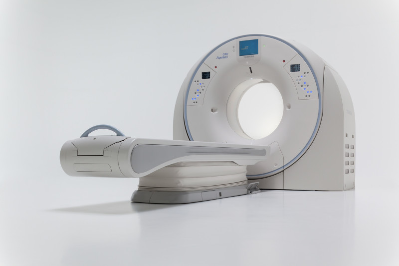
Toshiba Medical's Aquilion ONE -- GENESIS Edition premium CT
The cost-effective side of premium CTs
At last year’s Radiological Society of North America annual meeting, Toshiba Medical introduced its new Aquilion ONE / GENESIS Edition premium CT. The system filters the X-ray beam in a way that reduces dose and improves image quality.
Its PUREViSION Optics, PUREViSION Detector and FIRST reconstruction technology improve high contrast spacial resolution up to 129 percent while reducing dose by about 85 percent compared to the Aquilion ONE ViSION Edition.
Reconstruction times have been improved by up to 80 images per second and FIRST reconstruction is achieved in as little as three minutes per 320 images.
Three systems have been installed to date – the first at the National Institutes of Health and the other two at St. Cloud Hospital in Minnesota and Cincinnati Children’s Hospital Medical Center.
Tim Nicholson, senior manager of CT market development at Toshiba Medical, says that customers are willing to purchase premium CT systems if they can leverage them to improve patient outcomes and reduce costs.
“We hear of value-based medicine and we know radiology wants to show value,” he adds. “Customers are asking for something that proves you can use that technology and bring value to the patient's care and the institution that’s using it.”
In May, Toshiba Medical and the Stroke Care Center (SCC) at the Gates Vascular Institute in Buffalo, N.Y., published the results of their multi-year study on the economic impact of using the previous generation Aquilion ONE CT to diagnose and treat acute stroke.
The researchers gathered inpatient baseline data before the CT was installed between July and September 2007. Once the CT was installed, the 2009 to 2012 data revealed a significant increase in using CT-based protocols for stroke diagnoses, which translated into a reduction in health care costs and better patient outcomes.
Within the first five years after the Aquilion ONE was installed, SCC raked in more than $5.4 million in cost savings. That largely was due to reduced length of stays, less outpatient services and complications and lower readmission rates.
Toshiba Medical believes that any health care facility with a dedicated stroke program can reap the same cost savings.
More affordable options
"Health providers looking to enhance their CT capabilities may be faced with challenges such as reimbursement cuts, patient populations with low ability to pay or inefficiencies related to growth and expansion," says Philips' Gudapakkam.
In response to these trends, Philips introduced its Access CT system in March. The system was specifically designed for health care organizations that want to establish or improve their CT imaging capabilities at an accessible cost.
The iDose technology helps to reduce the need for tube replacement, which is the highest recurring cost associated with CT ownership. It's a reconstruction algorithm that reduces exposure time and extends tube life.
The iFlow console workflow platform offers features that simplify and automate the technologists’ routine, which helps them achieve consistently high image quality. It also helps the providers maintain and increase their referral base.
"[Access CT] expanded our capabilities into emerging markets and for cost-conscious health care organizations," says Gudapakkam. "As health care needs increase across the globe, many health care organizations are increasingly challenged to efficiently diagnose and treat a greater number of patients."
In April, Siemens received FDA clearance for its lower cost of ownership SOMATOM go. CT platform, which includes the SOMATOM go.Now and SOMATOM go.Up CT systems. It was designed as a single-room concept so facilities can save on installation costs and it has a smaller footprint compared to the company’s other CT platforms.
It’s primarily for routine CT work including head, chest, abdomen and pelvic exams, which make up 50 to 60 percent of CT exams. It’s ideal for community hospitals, critical access hospitals as well as outpatient imaging centers.
Siemens reported that the first few installations were underway in July and the company predicted that by the end of August approximately five would be installed in the U.S.
Personalized imaging for RT planning
The invention of CT played a crucial role in the development of radiation therapy planning. Other modalities like MR and PET are used secondarily in the treatment planning process, but CT remains the only 3-D imaging modality used for dose calculation, according to an article published in the journal BioMed Research International.
The newest CT simulator for radiation therapy to hit the market was Siemens' SOMATOM Confidence RT Pro CT system in December. Standard radiation therapy is built on 120 kV CT images, but this system can image patients at any kV setting.
“On the imaging side, it remains as a one-size-fits-all approach," says Hanno Dotzel, vice president of radiation oncology at Siemens. "The SOMATOM Confidence RT Pro in that regard is a game-changer because it allows for an image acquisition that is optimized for the individual size and anatomy of the patient."
The system's DirectDensity technology enables the technologist to optimize the scanner based on each individual patient, including bariatric patients and children.
“In radiology you would always optimize the scanner so you could get the best possible image quality, but in radiation oncology, there was always this workflow hurdle because for the dosimetry workflow, you need to have a calibration curve to tell you the electronic density," says Dotzel.
The SOMATOM Confidence RT Pro allows the technologists to freely choose a scan parameter without having to worry about the dosimetry workflow. That's because it's equipped with a technology that ensures they get the necessary information regardless of the kV settings.
Philips was the first to the CT simulator market with its Philips CT Big Bore scanner. GE followed with its Discover RT CT system, which was introduced at the American Society for Radiation Oncology meeting in November 2015.
Low-dose CT lung cancer screening
The National Lung Screening Trial, which was published in the New England Journal of Medicine in 2011, was the first to shine a light on the value of low-dose CT lung cancer screening.
The trial included 53,000 healthy older adults with a history of heavy smoking who were cared for at 33 different facilities. Half were screened with LDCT and the other half with X-ray and the results demonstrated that LDCT lowered the morality rate by 20 percent.
Members of Congress as well as a number of organizations joined forces in 2014 to push for Medicare coverage of LDCT lung cancer screening.
The moment the industry was waiting for came on Feb. 5, 2015, when Medicare issued its final decision on LDCT lung cancer screening. The agency decided that there was sufficient evidence to cover screening for individuals considered to be at high risk.
Those individuals are between the ages of 55 and 77 with no current signs or symptoms of lung cancer. They must have a smoking history of at least 30 pack-years or be current or former smokers who quit within the last 15 years.
GE was the first to get the FDA nod for LDCT lung cancer screening in August 2015. The screening protocol can be implemented on any of its 64-slice or greater CT scanners and most of its 16-slice systems.
In November 2015, the FDA cleared Siemens' SOMATOM CT systems for LDCT lung cancer screening, which includes the 16-slice SOMATOM Scope and ultra-premium SOMATOM Force.
In April 2016, Philips CT and PET/CT models were approved by the FDA for LDCT lung cancer screening. All 27 models can also be used for dose management and image data sharing and analytics through the Philips IntelliSpace Portal.
Despite the initial hype, the adoption of LDCT lung cancer screening has been relatively slow. According to Ken Denison, molecular imaging and CT dose leader at GE, that's due to the challenges associated with getting CMS reimbursement.
"They have to prove they had a smoking cessation conversation and risk-benefit conversation with the patient," he adds. "That whole process is pretty challenging to implement from scratch."
In addition, general practitioners have mixed views on its value. Given that they're the ones that primarily drive the conversation to recruit smokers and former smokers into a program, that presents a challenge as well.
There is also reluctance expressed by patients. Denison explains that if patients don't feel like they have a problem, they don't see the point in getting screened.
In an attempt to change that, Siemens is working with several providers that are interested in deploying an RV vehicle equipped with a 16-slice CT that can travel throughout their service area and deliver lung cancer screening directly to the patients.
“People are definitely ramping up, but it’s going much more slowly than it could be," says Denison. "But even mammography back in the day ramped up more slowly than it could have. Change takes time.”