New approach examines variety of
components to determine radiation
exposure and preserve image quality
components to determine radiation
exposure and preserve image quality
Determining pediatric radiation dose and CT image quality with quantitative method
September 13, 2017
by John R. Fischer, Senior Reporter
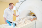
A new technique may ensure proper amounts in radiation exposure and better image quality across a range of different sized pediatric patients during CT.
Researchers at Duke University Medical Center and Cleveland Clinic have developed a quantitative method that can assess a variety of components for determining the appropriate amount of radiation to administer while still preserving high image quality during CT exams, based on an individual patient’s body type. A study on the approach was published in the Journal of Medical Imaging.
“It’s a composite of different elements, all of which are important, but to date, they’ve not really all been put together systematically,” Dr. Donald P. Frush, a professor of radiology and pediatrics at Duke University and an author of the study, told HCB News. “You make decisions based on the size of the patient but once you have a particular sized patient who’s 10 years old and it’s an abdomen pelvis CT, you just do it the same way each time that you’re dealing with a 10-year-old patient. That’s really not what we want to do. We want to make individual patient adjustments, asking ‘What’s the body type of this patient, what are we needing to see, and what risk profile will be appropriate?’”
Current methods for assessing proper radiation dose while keeping image quality high may substantially vary from one practice to another. Even within a practice, the method used is based on what the individual radiologist deems as acceptable criteria for determining these aspects.
The proposed approach creates a more systematic order that takes into consideration the effects of many components, such as age, size/weight and gender and the CT settings that impact dose and image quality. The dose estimations are based on organ doses derived from previous published models by these investigators.
The radiologist, in designing indication-based protocols, can then determine how much to maintain the same image quality across all sized pediatric patients and make modifications to ensure consistent radiation exposure, while still operating within an acceptable range of image quality.
The approach is based on two foundational studies in which organ doses, effective dose and a risk index were determined for nine groups of pediatric patients based on age and size. With the addition of noise and simulated lung nodule lesions, nodule detection accuracy was determined with the results of the accuracy-dose relationship used to guide selection of individual scan parameters for each category of patients, for this indication.
Based on the results, the technique could be used as a guide for determining appropriate development of medical imaging protocols across pediatric and adult populations for a variety of imaging modalities beyond CT.
Frush cautions though that using the approach for each specific indication and other modalities requires much work.
“I think if there were further development of this template, where the various components of image quality based on indication and the individual patient factors could be added (and that takes some additional study), then that could become a tool for clinical use,” he said. “Again, I think an ultimate application would be that vendors could work with research partners and implement this on their equipment, so that someone would just be able to select the built-in protocol designed for a consistent approach to diagnostic value (mindful of risk) based on the indication and with the individual considerations of the child on the scanner.”
Following more research, the approach may be proven as a potential way for more efficient and enhanced protocol development, review, and auditing, as well as reducing exam time-saving costs from an operational standpoint.