
Siemens Healthineers Magnetom
Vida 3T MR system
Vida 3T MR system
MR providers seek faster scans and simplified workflow
October 02, 2017
by Lauren Dubinsky, Senior Reporter
It’s no secret that health care organizations are under pressure to do more with less.
When new MR systems appear on the market that can dramatically reduce scan time – providers notice.
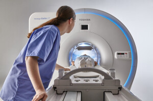
“It’s a very fast system,” says Dr. Mark Winkler of Steinberg Diagnostic Medical Imaging in Las Vegas, describing his facility’s Vantage Galan 3T MR from Toshiba Medical. “Our maximum protocol length is two minutes, and that’s without compromising slice thickness or spatial resolution.”
Compared to other systems on the market that can take twice as long, this scanner creates new opportunities for Winkler and his colleagues to improve patient throughput or spend time on other important tasks.
He uses the scanner for typical neurology and musculoskeletal exams, as well as basic to advanced body oncology work. It’s also his go-to system for advanced applications like cerebrospinal fluid flow studies and brain/body spectroscopy.
“One of the problems with 3T is that the advanced applications for sequences can get very noisy,” says Winkler. The Vantage Galan 3T utilizes Toshiba’s Pianissimo Zen technology to reduce that noise and, as a result, cause less anxiety and claustrophobia for the person inside the magnet.
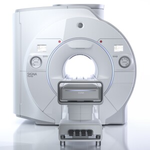
GE Healthcare’s new SIGNA Premier wide-bore 3T MR, which received FDA clearance in August, is also raising the bar on acquisition time.
“Patients are often sick, uncomfortable, or anxious for answers — it’s not fun to be in the scanner,” says Eric Stahre, president and CEO of MR at GE. “You want to get in and get out. With an acquisition that’s eight times faster, that benefits everybody: less movement from the patient and improved productivity on the scanner.”
The SIGNA Premier, which can perform routine brain exams in less than five minutes, was the product of a four-year partnership GE had with the National Football League and research institutions. The goal was to design new imaging tools for detecting biomarkers that could potentially diagnose mild traumatic brain injury.
The system uses HyperSense technology, which is part of GE’s HyperWorks application suite, and also boasts high-performance, SuperG gradients that can switch faster than previous systems and have higher strength – all factors that contribute to the speedy scan time.
The University of Wisconsin-Madison acquired the SIGNA Premier through a research agreement with GE. It has scanned between 120 and 150 healthy volunteers and 230 clinical patients as part of an institutional review board evaluation.
Another advantage of the SuperG gradients is that they can be used with diffusion-weighted imaging (DWI) as a powerful tool for detecting stroke and characterizing cancer.
“DWI is very demanding on the system and requires high-performance gradients,” says Dr. Scott Reeder, chief of MR imaging and vice chairman of research at the university. “We have noticed significant improvements in performance for DWI, largely because of the improved gradient performance.”
In February, Siemens Healthineers got the FDA nod for its Compressed Sensing MR acceleration technology, which is available on its MAGNETOM Aera 1.5T and MAGNETOM Skyra 3T systems. With this technology, cardiac cine imaging can be completed in 16 seconds compared to the conventional four minutes.
Rather than having to hold their breath 10 to 14 times, patients are able to breathe freely during the exam. This eliminates motion artifacts that are created by breathing and heartbeats, which is especially useful for older, critically ill and pediatric patients.
“If you’re performing the exam fast enough that the patient doesn’t have to hold their breath, you’re expanding the clinical capabilities and the patient population you can reach,” says Chris Farischon, MR product manager at Siemens. “Patients who may not have been able to have a cardiac MR exam previously could now potentially have one of those exams.”
Faster scan time is not just something for research and academic facilities. In February, Siemens got FDA clearance for its MAGNETOM Sempra, a system that can perform brain scans in 10 minutes and is being touted as the manufacturer’s “most cost-effective” 1.5 T MR scanner.

Is there a strong demand for 3T systems?
While speeding up scan time, new, high-capability, budget-conscious solutions like the MAGNETOM Sempra may be changing the way providers think about what they should be shopping for.
“If an imaging center is replacing an older system and they don’t have a need for a 70-centimeter bore because they’re looking for a low-cost solution, that’s exactly what Sempra was meant to address,” says Farischon. “They’re low total-cost-of-ownership systems from installation to operation.”
Its Day optimizing throughput (Dot) workflow engines for brain, spine and large joint exams adjust the clinical cases for each patient. Those cases comprise up to 75 percent of the average MR imaging volume, which means most exams can be conducted faster and repeat scans can be avoided more often.
At the 2016 RSNA meeting, Siemens said that motion alone can cost $100,000 per year due to the number of re-scans that are required.
If a 1.5T system can provide this level of performance, is there still demand for 3T systems?
In 2013, 60 percent of ECRI’s members were interested in 1.5T systems while 35 percent were interested in 3T systems. But in 2016, the interest in 1.5T systems rose to 68 percent and interest in 3T systems dropped to 30 percent.
According to Jason Launders, director of operations for the Health Devices Group at ECRI, significant financial pressure has resulted in hospitals not spending as freely as they once did.
Launders explains that there is about a $500,000 price difference between 1.5T and 3T systems. The latter provides better image quality and comes with more tools, but 1.5T technology has advanced in recent years.
But manufacturers may view the market differently, such as GE, which reports a decline in facilities purchasing new 1.5T systems.
“We still see strength in 3T versus 1.5T. I think the shift there is happening because of the number of advanced MR features that are growing,” says Stahre.
“If there is a decline,” he adds, “it’s in new 1.5T systems versus refurbished systems.”
Based on data from the National Electrical Manufacturers Association (NEMA), Stahre says that the ratio of new 1.5T to 3T system purchases is 2 to 1, and that the 3T side is continuing to grow.
Making neurology applications more user-friendly
The global MR market is expected to reach approximately $5.6 billion by 2025, according to a Grand View Research market report from January. MR for neural and brain disorders dominated the market, since there is no alternate scanning tool available for those applications.
Another survey, published by TMTG last year, found that 70 percent of radiologists report having difficulty using their existing neuro-diagnostic tools because they lack effective imaging and visualization techniques.
To assist with this, Philips launched three new MR-based neurology software applications for its Ingenia digital MR systems at RSNA 2016. These tools are designed to assist radiologists in making more definitive diagnoses.
“MR has traditionally been used for soft tissue characterization for oncology and vascular disease in the brain,” says Martijn Hartjes, senior director and head of global MR marketing at Philips. “We see new areas in neuro being explored, like neurodegenerative types of diseases, such as Alzheimer's [disease], [traumatic brain injury] and neuropsychiatry."
The new suite of neuro-diagnostic applications are equipped with unique features like MultiBand SENSE, which allows for the simultaneous acquisition of multiple slices of the brain during fMRI exams. It also gives radiologists the option to increase coverage or resolution without prolonging the scan.
The Black Blood Imaging feature reduces the blood signal, which generates a high-resolution 3-D brain image, and the 4-D-TRANCE feature provides contrast-free 4-D imaging of the brain vascular anatomy.
“The main driver behind [developing the suite] was that we saw that a lot of radiologists still had difficulty answering the more complex questions in the domain of neurology,” says Hartjes. “We did some assessments of the main areas and built a suite around that.”
Progress toward personalized MR
As value-based care becomes more prevalent, personalized medicine is playing a bigger role in managing population health. But treatment is only one side of the coin — diagnostics is on the other side.
ECRI’s Launders explains that it’s been a gradual trend over the last 10 to 15 years. It started with dedicated coils and then general purpose and matrix coils were introduced, which are easier to position on patients.
In March, Siemens launched its Magnetom Vida 3T MR at the University Hospital Tübingen in Germany. It’s the first system equipped with the company’s BioMatrix technology, which adjusts the system to fit the anatomy and physiology of each patient.
The system uses intelligent body models to automatically move the patient table into the correct scan position. The BioMatrix sensors inside the table track patients’ respiratory patterns to assess if they can hold their breath during the exam.
The BioMatrix tuners improve the quality and reproducibility of whole-body diffusion, which helps avoid repeat scans. It also controls the scan parameters in real time in order to stay consistent with the patient’s anatomy and avoid distortions that make the diffusion image non-diagnostic.
"Siemens' introduction looks like very exciting technology, but it’s going to take time before you see it everywhere,” says Launders.
He adds that it was the same case with wide-bore technology when Toshiba Medical introduced it 10 years ago, and technology that makes the exam quieter. All new systems now feature both of those technologies.
New coil introductions
For the Magnetom Vida, Siemens also introduced new coils, including ultra-flex coils for shoulders, knees and other parts of the body. They are high density coils made of memory foam material for easier use.
The company also introduced two dedicated coils. The dedicated knee coil has a larger inner aperture compared to previous generation coils, so that it can fit larger patients.
“All of this technology is designed to help diagnose the patient’s individual condition,” says Farischon. “As a single thing, you may not be able to point to personalized medicine, but with all of these technologies as a whole, you can clearly see that we are moving in that direction.”
At RSNA 2016, GE unveiled its AIR Coil surface coil for the SIGNA Premier and it’s two-thirds lighter than standard coils. It’s similar to placing a blanket on the patient, which makes it easier for the technologist to maneuver it to conform to the patient’s body.
"When you're a patient in the bore and the technologist is putting all this body armor on top of you to provide a good, local antenna to receive the MR signal, it can be bulky and heavy: like body armor to the patient and not fun for the technologist to move around,” says GE’s Stahre.
In addition to being lighter, the AIR Coil can also receive a stronger MR signal because it’s next to the body.
Stahre says that the AIR Coil is a major breakthrough for the industry and that more technology like this will emerge over the next couple of years.
Helium-free MR
GE is working on another industry-first technology that it also introduced at RSNA 2016. The Freelium magnet technology is designed to use 1 percent of liquid helium compared to conventional MR magnets — instead of 2,000 liters of helium, it uses about 20 liters.
With the technology, a hospital wouldn’t need to install the extensive venting that’s usually required when putting a magnet in a separate building for a newly constructed room. The Freelium magnet also doesn’t have to be refilled during transportation or even throughout its life cycle.
GE has a prototype in operation at the Mayo Clinic in Rochester, Minn., for research purposes. It has not been cleared by the FDA or any other regulatory body, and is under development.
"Helium is a limited resource and it is becoming expensive,” says ECRI’s Launders. “If you can eliminate the need to refill helium, that’s useful."
New MR systems come with zero boil-off technology, which recondenses the helium with a cold head. The issue is that it uses a lot of energy, and if the power is lost, then the facility loses its helium.
Launders sees the benefits that the Freelium technology would have, but he says GE has to prove that it works before the industry can call it a breakthrough.
When new MR systems appear on the market that can dramatically reduce scan time – providers notice.
“It’s a very fast system,” says Dr. Mark Winkler of Steinberg Diagnostic Medical Imaging in Las Vegas, describing his facility’s Vantage Galan 3T MR from Toshiba Medical. “Our maximum protocol length is two minutes, and that’s without compromising slice thickness or spatial resolution.”
Compared to other systems on the market that can take twice as long, this scanner creates new opportunities for Winkler and his colleagues to improve patient throughput or spend time on other important tasks.
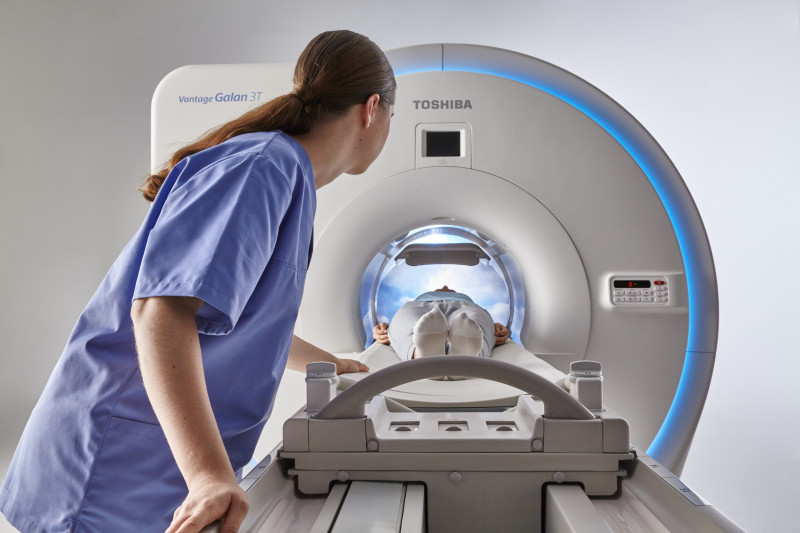
Toshiba Medical Systems Corporation's
Vantage Galan 3T MR system
Vantage Galan 3T MR system
He uses the scanner for typical neurology and musculoskeletal exams, as well as basic to advanced body oncology work. It’s also his go-to system for advanced applications like cerebrospinal fluid flow studies and brain/body spectroscopy.
“One of the problems with 3T is that the advanced applications for sequences can get very noisy,” says Winkler. The Vantage Galan 3T utilizes Toshiba’s Pianissimo Zen technology to reduce that noise and, as a result, cause less anxiety and claustrophobia for the person inside the magnet.
GE Healthcare’s new SIGNA Premier wide-bore 3T MR, which received FDA clearance in August, is also raising the bar on acquisition time.
“Patients are often sick, uncomfortable, or anxious for answers — it’s not fun to be in the scanner,” says Eric Stahre, president and CEO of MR at GE. “You want to get in and get out. With an acquisition that’s eight times faster, that benefits everybody: less movement from the patient and improved productivity on the scanner.”
The SIGNA Premier, which can perform routine brain exams in less than five minutes, was the product of a four-year partnership GE had with the National Football League and research institutions. The goal was to design new imaging tools for detecting biomarkers that could potentially diagnose mild traumatic brain injury.
The system uses HyperSense technology, which is part of GE’s HyperWorks application suite, and also boasts high-performance, SuperG gradients that can switch faster than previous systems and have higher strength – all factors that contribute to the speedy scan time.
The University of Wisconsin-Madison acquired the SIGNA Premier through a research agreement with GE. It has scanned between 120 and 150 healthy volunteers and 230 clinical patients as part of an institutional review board evaluation.
Another advantage of the SuperG gradients is that they can be used with diffusion-weighted imaging (DWI) as a powerful tool for detecting stroke and characterizing cancer.
“DWI is very demanding on the system and requires high-performance gradients,” says Dr. Scott Reeder, chief of MR imaging and vice chairman of research at the university. “We have noticed significant improvements in performance for DWI, largely because of the improved gradient performance.”
In February, Siemens Healthineers got the FDA nod for its Compressed Sensing MR acceleration technology, which is available on its MAGNETOM Aera 1.5T and MAGNETOM Skyra 3T systems. With this technology, cardiac cine imaging can be completed in 16 seconds compared to the conventional four minutes.
Rather than having to hold their breath 10 to 14 times, patients are able to breathe freely during the exam. This eliminates motion artifacts that are created by breathing and heartbeats, which is especially useful for older, critically ill and pediatric patients.
“If you’re performing the exam fast enough that the patient doesn’t have to hold their breath, you’re expanding the clinical capabilities and the patient population you can reach,” says Chris Farischon, MR product manager at Siemens. “Patients who may not have been able to have a cardiac MR exam previously could now potentially have one of those exams.”
Faster scan time is not just something for research and academic facilities. In February, Siemens got FDA clearance for its MAGNETOM Sempra, a system that can perform brain scans in 10 minutes and is being touted as the manufacturer’s “most cost-effective” 1.5 T MR scanner.
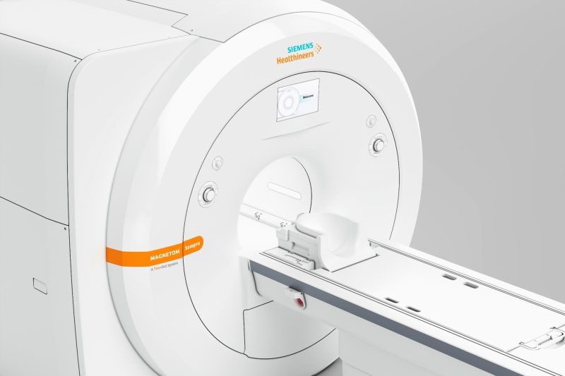
Siemens Healthineers' MAGNETOM Sempra 1.5T MR system
Is there a strong demand for 3T systems?
While speeding up scan time, new, high-capability, budget-conscious solutions like the MAGNETOM Sempra may be changing the way providers think about what they should be shopping for.
“If an imaging center is replacing an older system and they don’t have a need for a 70-centimeter bore because they’re looking for a low-cost solution, that’s exactly what Sempra was meant to address,” says Farischon. “They’re low total-cost-of-ownership systems from installation to operation.”
Its Day optimizing throughput (Dot) workflow engines for brain, spine and large joint exams adjust the clinical cases for each patient. Those cases comprise up to 75 percent of the average MR imaging volume, which means most exams can be conducted faster and repeat scans can be avoided more often.
At the 2016 RSNA meeting, Siemens said that motion alone can cost $100,000 per year due to the number of re-scans that are required.
If a 1.5T system can provide this level of performance, is there still demand for 3T systems?
In 2013, 60 percent of ECRI’s members were interested in 1.5T systems while 35 percent were interested in 3T systems. But in 2016, the interest in 1.5T systems rose to 68 percent and interest in 3T systems dropped to 30 percent.
According to Jason Launders, director of operations for the Health Devices Group at ECRI, significant financial pressure has resulted in hospitals not spending as freely as they once did.
Launders explains that there is about a $500,000 price difference between 1.5T and 3T systems. The latter provides better image quality and comes with more tools, but 1.5T technology has advanced in recent years.
But manufacturers may view the market differently, such as GE, which reports a decline in facilities purchasing new 1.5T systems.
“We still see strength in 3T versus 1.5T. I think the shift there is happening because of the number of advanced MR features that are growing,” says Stahre.
“If there is a decline,” he adds, “it’s in new 1.5T systems versus refurbished systems.”
Based on data from the National Electrical Manufacturers Association (NEMA), Stahre says that the ratio of new 1.5T to 3T system purchases is 2 to 1, and that the 3T side is continuing to grow.
Making neurology applications more user-friendly
The global MR market is expected to reach approximately $5.6 billion by 2025, according to a Grand View Research market report from January. MR for neural and brain disorders dominated the market, since there is no alternate scanning tool available for those applications.
Another survey, published by TMTG last year, found that 70 percent of radiologists report having difficulty using their existing neuro-diagnostic tools because they lack effective imaging and visualization techniques.
To assist with this, Philips launched three new MR-based neurology software applications for its Ingenia digital MR systems at RSNA 2016. These tools are designed to assist radiologists in making more definitive diagnoses.
“MR has traditionally been used for soft tissue characterization for oncology and vascular disease in the brain,” says Martijn Hartjes, senior director and head of global MR marketing at Philips. “We see new areas in neuro being explored, like neurodegenerative types of diseases, such as Alzheimer's [disease], [traumatic brain injury] and neuropsychiatry."
The new suite of neuro-diagnostic applications are equipped with unique features like MultiBand SENSE, which allows for the simultaneous acquisition of multiple slices of the brain during fMRI exams. It also gives radiologists the option to increase coverage or resolution without prolonging the scan.
The Black Blood Imaging feature reduces the blood signal, which generates a high-resolution 3-D brain image, and the 4-D-TRANCE feature provides contrast-free 4-D imaging of the brain vascular anatomy.
“The main driver behind [developing the suite] was that we saw that a lot of radiologists still had difficulty answering the more complex questions in the domain of neurology,” says Hartjes. “We did some assessments of the main areas and built a suite around that.”
Progress toward personalized MR
As value-based care becomes more prevalent, personalized medicine is playing a bigger role in managing population health. But treatment is only one side of the coin — diagnostics is on the other side.
ECRI’s Launders explains that it’s been a gradual trend over the last 10 to 15 years. It started with dedicated coils and then general purpose and matrix coils were introduced, which are easier to position on patients.
In March, Siemens launched its Magnetom Vida 3T MR at the University Hospital Tübingen in Germany. It’s the first system equipped with the company’s BioMatrix technology, which adjusts the system to fit the anatomy and physiology of each patient.
The system uses intelligent body models to automatically move the patient table into the correct scan position. The BioMatrix sensors inside the table track patients’ respiratory patterns to assess if they can hold their breath during the exam.
The BioMatrix tuners improve the quality and reproducibility of whole-body diffusion, which helps avoid repeat scans. It also controls the scan parameters in real time in order to stay consistent with the patient’s anatomy and avoid distortions that make the diffusion image non-diagnostic.
"Siemens' introduction looks like very exciting technology, but it’s going to take time before you see it everywhere,” says Launders.
He adds that it was the same case with wide-bore technology when Toshiba Medical introduced it 10 years ago, and technology that makes the exam quieter. All new systems now feature both of those technologies.
New coil introductions
For the Magnetom Vida, Siemens also introduced new coils, including ultra-flex coils for shoulders, knees and other parts of the body. They are high density coils made of memory foam material for easier use.
The company also introduced two dedicated coils. The dedicated knee coil has a larger inner aperture compared to previous generation coils, so that it can fit larger patients.
“All of this technology is designed to help diagnose the patient’s individual condition,” says Farischon. “As a single thing, you may not be able to point to personalized medicine, but with all of these technologies as a whole, you can clearly see that we are moving in that direction.”
At RSNA 2016, GE unveiled its AIR Coil surface coil for the SIGNA Premier and it’s two-thirds lighter than standard coils. It’s similar to placing a blanket on the patient, which makes it easier for the technologist to maneuver it to conform to the patient’s body.
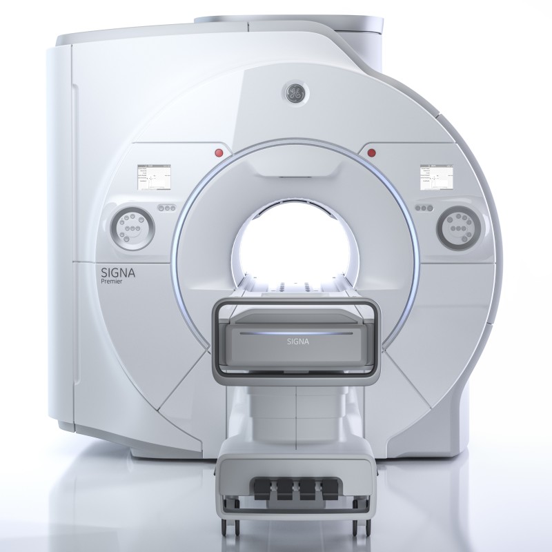
GE Healthcare's SIGNA Premier 3T MR system
"When you're a patient in the bore and the technologist is putting all this body armor on top of you to provide a good, local antenna to receive the MR signal, it can be bulky and heavy: like body armor to the patient and not fun for the technologist to move around,” says GE’s Stahre.
In addition to being lighter, the AIR Coil can also receive a stronger MR signal because it’s next to the body.
Stahre says that the AIR Coil is a major breakthrough for the industry and that more technology like this will emerge over the next couple of years.
Helium-free MR
GE is working on another industry-first technology that it also introduced at RSNA 2016. The Freelium magnet technology is designed to use 1 percent of liquid helium compared to conventional MR magnets — instead of 2,000 liters of helium, it uses about 20 liters.
With the technology, a hospital wouldn’t need to install the extensive venting that’s usually required when putting a magnet in a separate building for a newly constructed room. The Freelium magnet also doesn’t have to be refilled during transportation or even throughout its life cycle.
GE has a prototype in operation at the Mayo Clinic in Rochester, Minn., for research purposes. It has not been cleared by the FDA or any other regulatory body, and is under development.
"Helium is a limited resource and it is becoming expensive,” says ECRI’s Launders. “If you can eliminate the need to refill helium, that’s useful."
New MR systems come with zero boil-off technology, which recondenses the helium with a cold head. The issue is that it uses a lot of energy, and if the power is lost, then the facility loses its helium.
Launders sees the benefits that the Freelium technology would have, but he says GE has to prove that it works before the industry can call it a breakthrough.