A new all-optical ultrasound may soon
enable simultaneous imaging for
MR and ultrasound
enable simultaneous imaging for
MR and ultrasound
All-optical ultrasound could pave way for combined MR-ultrasound imaging
July 13, 2018
by John R. Fischer, Senior Reporter
Simultaneous ultrasound and MR imaging has, up until this point, been impossible due to the dangers associated with exposing electronic components in ultrasound systems to the magnetic influence of MR.
But such a task may soon be a reality with the introduction of an all-optical ultrasound, the first to provide video-rate, real-time 2D imaging of biological tissue at a frame rate of up to three orders of magnitude faster than current optical systems.
“Advantages expected for applications range from routine bedside exams to image guidance of minimally invasive procedures,” Dr. Erwin J. Alles, a senior research associate at the department of medical physics and biomedical engineering at University College London, told HCB News. “The later application especially, where devices are considered disposable, will benefit greatly from the reduced costs of the all-optical technology and from MR compatibility.”
Conventional ultrasounds are composed of electronic transducers that transmit high-frequency sound waves into tissues, which send back reflections that are used by a computer to construct images.
However, the presence of these components prevents simultaneous imaging with MR platforms and creates challenges in the manufacturing of small ultrasounds for internal use, resulting in systems with large, handheld probes pressed against the skin. While high-resolution minimally invasive ultrasound probes are available, many are unaffordable for routine clinical use.
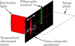
Optical components, in contrast, can be easily miniaturized and utilize probes that are significantly less expensive to produce. Its scanning mirror controls the transmission of waves generated by pulsed laser light and received by fiber optic sensors, increasing image quality and enabling image acquisition to take place across different modes, such as 2D and 3D imaging, on a single device.
This eliminates the need for highly specialized electronic ultrasound-based probes which can be highly disruptive and extend procedure time and risk to patients.
The lack of electrical parts allows the system to be safely used alongside MR scanners, providing clinicians with comprehensive pictures depicting tissues around areas of interest such as tumors or blood.
The generation of all-optical images has traditionally been a time-consuming process, relying on multiple optical source locations for the retrieval of data that is then combined to create a reconstruction of the area being imaged.
To overcome this, researchers increased speed with the addition of a new imaging paradigm, new optical ultrasound generating materials, optimized ultrasound source geometries, and a highly sensitive fiber optic ultrasound detector.
Optical systems also produce larger bandwidths of sound than electronic-based ones, making them more versatile. By manipulating the light sources, researchers generated both low-frequency ultrasound for greater penetration into tissue and high-frequency ultrasound for higher resolution of images at shallower depth.
The team tested the protype system by imaging a deceased zebrafish and a pig artery that was manipulated to emulate the dynamics of pulsing blood. Compared to an electronic high-frequency ultrasound, the system demonstrated a sustained frame rate of 15 Hertz, a dynamic range of 30 decibels, a penetration depth of six millimeters and a resolution of 75 by 100 micrometers.
Alles says the implementation of the scanner is expected to provide access to quality and affordable imaging and aid providers in administering greater and more valuable care to patients.
The potential for reduced costs of minimally invasive imaging probes will allow for more widespread application of high-quality image guidance during minimally invasive procedures,” he said. “Such minimally invasive procedures improve patient outcomes, reduce procedure time and complication rates, and usually reduce costs. Adding all-optical ultrasound to minimally invasive probes will result in improved visualization and guidance of the procedure, and will help overcome barriers preventing widespread use of minimally invasive procedures.”
Researchers are currently working to adapt the technology for clinical use with the development of a long, flexible imaging probe that enables freehand operation, and miniaturized versions for endoscopic applications.
Plans for human studies are currently underway to validate and eventually commercialize the system for clinical use.
Commercialization for preclinical and non-biological contexts is also anticipated.
But such a task may soon be a reality with the introduction of an all-optical ultrasound, the first to provide video-rate, real-time 2D imaging of biological tissue at a frame rate of up to three orders of magnitude faster than current optical systems.
“Advantages expected for applications range from routine bedside exams to image guidance of minimally invasive procedures,” Dr. Erwin J. Alles, a senior research associate at the department of medical physics and biomedical engineering at University College London, told HCB News. “The later application especially, where devices are considered disposable, will benefit greatly from the reduced costs of the all-optical technology and from MR compatibility.”
Conventional ultrasounds are composed of electronic transducers that transmit high-frequency sound waves into tissues, which send back reflections that are used by a computer to construct images.
However, the presence of these components prevents simultaneous imaging with MR platforms and creates challenges in the manufacturing of small ultrasounds for internal use, resulting in systems with large, handheld probes pressed against the skin. While high-resolution minimally invasive ultrasound probes are available, many are unaffordable for routine clinical use.
Optical components, in contrast, can be easily miniaturized and utilize probes that are significantly less expensive to produce. Its scanning mirror controls the transmission of waves generated by pulsed laser light and received by fiber optic sensors, increasing image quality and enabling image acquisition to take place across different modes, such as 2D and 3D imaging, on a single device.
This eliminates the need for highly specialized electronic ultrasound-based probes which can be highly disruptive and extend procedure time and risk to patients.
The lack of electrical parts allows the system to be safely used alongside MR scanners, providing clinicians with comprehensive pictures depicting tissues around areas of interest such as tumors or blood.
The generation of all-optical images has traditionally been a time-consuming process, relying on multiple optical source locations for the retrieval of data that is then combined to create a reconstruction of the area being imaged.
To overcome this, researchers increased speed with the addition of a new imaging paradigm, new optical ultrasound generating materials, optimized ultrasound source geometries, and a highly sensitive fiber optic ultrasound detector.
Optical systems also produce larger bandwidths of sound than electronic-based ones, making them more versatile. By manipulating the light sources, researchers generated both low-frequency ultrasound for greater penetration into tissue and high-frequency ultrasound for higher resolution of images at shallower depth.
The team tested the protype system by imaging a deceased zebrafish and a pig artery that was manipulated to emulate the dynamics of pulsing blood. Compared to an electronic high-frequency ultrasound, the system demonstrated a sustained frame rate of 15 Hertz, a dynamic range of 30 decibels, a penetration depth of six millimeters and a resolution of 75 by 100 micrometers.
Alles says the implementation of the scanner is expected to provide access to quality and affordable imaging and aid providers in administering greater and more valuable care to patients.
The potential for reduced costs of minimally invasive imaging probes will allow for more widespread application of high-quality image guidance during minimally invasive procedures,” he said. “Such minimally invasive procedures improve patient outcomes, reduce procedure time and complication rates, and usually reduce costs. Adding all-optical ultrasound to minimally invasive probes will result in improved visualization and guidance of the procedure, and will help overcome barriers preventing widespread use of minimally invasive procedures.”
Researchers are currently working to adapt the technology for clinical use with the development of a long, flexible imaging probe that enables freehand operation, and miniaturized versions for endoscopic applications.
Plans for human studies are currently underway to validate and eventually commercialize the system for clinical use.
Commercialization for preclinical and non-biological contexts is also anticipated.