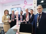
Q&A with Danilo Gennari
May 03, 2019
by Gus Iversen, Editor in Chief
DeHCA Light & Sound is a new form of breast cancer detection technology, but rather than visualize tumors it relies on angiogenesis to detect increases in deoxyhemoglobin. HealthCare Business News spoke to Danilo Gennari, an engineer and member of the Board of the Italian Association of Clinical Engineering (AIIC) who is the scientific director of the DeHCA Project, and principal creator of the device.
HCB News: Without getting too technical, what is angiogenesis and what is deoxyhemoglobin?
Danilo Gennari: Angiogenesis is the growth of new capillary blood vessels in the body; it is an important natural process used for healing and reproduction. The body controls angiogenesis by producing a precise balance of growth and inhibitory factors in healthy tissues. When this balance is disturbed, the result is either too much or too little angiogenesis. Abnormal blood vessel growth (abnormal angiogenesis), either excessive or insufficient, is now recognized as a “common denominator” underlying many deadly and debilitating conditions, including cancer, skin diseases, age-related blindness, diabetic ulcers, cardiovascular disease, stroke, and many others. The list of diseases that have angiogenesis as an underlying mechanism grows longer every year. The oxygen consumption in growing tumors is higher than in normal tissue and results in abnormally deoxygenated hemoglobin – abnormal concentration of deoxy-hemoglobin – in the blood vessels feeding the tumors.
HCB News: What is the relationships between deoxyhemoglobin and breast cancer?
DG: Neo-angiogenesis is the phenomenon by which tumor cells grow beyond one millimiter diameter approximately, developing their own circulation system to get fed. This is done mimicking the circulatory system of the healthy tissue nearby. As said, neo-angiogenetic blood vessels are characterized by abnormally high concentration of deoxy-hemoglobin. Consequently, abnormal concentration of deoxy-hemoglobin can be associated to the neo-angiogenesis at the root of the formation of new tumors.
HCB News: The system is primarily comprised of an LED light table and an ultrasound transducer. How do these two components work together to detect potential cancers?
DG: The DeHCA L&S idea is that, in early stage tumors, it is easier to find the tumor-feeding vascular network rather than the tumor itself, because the vascular network arises early in the disease, and results in vascular changes affecting vessels beyond the tumor itself, thus being earlier identifiable. Identification is achieved evaluating the DeHCA biomarker (Deoxy Hemoglobin Concentration Alteration) which represents the variation of concentration of deoxy-hemoglobin within the breast tissues and, as said previously, its abnormal values can be associated to the neo-angiogenesis at the root of the formation of new tumors.
With DeHCA L&S physicians perform an initial optical examination with the objective to identify and locate areas of likely lesions, through the evaluation of the DeHCA biomarker. If any suspect areas are identified, physicians proceed to an ultrasound examination to exactly locate and qualify the lesions and eventually support a guided biopsy.
The optical information is acquired illuminating the breast with bottom-up 640nm red light, emitted by the LED’s positioned in the LED table (LED Plate). 640nm is the wavelength at which healthy and pathologic tissues show the maximum difference in transparency, which is lower in case of tumor neo-angiogenesis. Low transparency areas have presence of pathologic vessels.
The ultrasound information is used to exactly locate the lesion within the areas identified by the optical scan. The ultrasound morphological images and optical functional images can be directly compared.
HCB News: Can DeHCA provide a definitive diagnosis or would a patient with increased deoxyhemoglobin be referred for more follow up imaging?
DG: With DeHCA L&S the diagnosis is made on the base of the ultrasound examination, while the optical examination is used as viewfinder. This procedure can be compared to what presently done with mammography and ultrasound. Therefore, we expect the DeHCA L&S diagnosis to be confirmed by biopsy, as in today’s clinical protocols.
HCB News: The system depends in part on a patented applicator that sort of vacuum seals onto the patients breast. What purpose does that serve and is it painful?
DG: The evaluation of the concentration of deoxy-hemoglobin must be done when blood circulation in capillaries is temporarily stopped. This is achieved through a mild pressure exercised by the Applicator. The Applicator is a simple pressure/vacuum mechanism, at first glance similar to a bra, that makes the external ambient pressure to be superior to the pressure inside. In this way the Applicator membrane collapses on the breast and, perfectly adhering to it, provokes its immobilization on LED Plate and the temporary blockage of blood circulation in the neo-angiogenetic capillary vessels.
The mild applied pressure is about 20mmHg, absolutely painless, very much less than pressure applied to the breast with mammography.
HCB News: Is there any indication that breast density impacts the visualization of deoxyhemoglobin?
DG: No. Breast density does not impact the visualization of deoxy-hemoglobin, because the 640nm red light is not stopped by glandular tissues.
HCB News: Who would be the ideal candidates to undergo DeHCA imaging?
DG: Mammography shows limited efficiency on dense breasts, which is not the case with DeHCA L&S. Therefore, the initial and ideal candidates to undergo DeHCA L&S examination are dense-breast women, typically young women today not considered by national screening plans. We should not forget that dense breast is considered a risk factor and characterizes almost all women in pre-menopausal status and about the 25% of women in post-menopausal status, with trend to increase with the increase of wellness. This percentage is even higher in most Asian countries.
Furthermore, breast glandular tissue has a high radiosensitivity to the ionizing radiations used with mammography, particularly in premenopausal women. DeHCA L&S does not emit any ionizing radiation.
HCB News: What stage of development is the system in?
DG: At present, we are validating the device in a two-site trial. Two DeHCA L&S are installed at the European Institute of Oncology (IEO) and at the S. Raffaele Hospital (HSR), both in Milan (Italy). In parallel, we are continuing testing DeHCA L&S on volunteers at our lab. We plan to go to market in 2020.
HCB News: In terms of lowering costs and improving outcomes, what does DeHCA Light & Sound fit into the healthcare ecosystem?
DG: The adoption of DeHCA L&S would lead to important savings in both direct and indirect costs of the healthcare ecosystem. This because the early detection ability will lead to less and lighter surgical interventions, softer radiant and drug therapies, reduced costs for physical rehabilitation and psychological support, shorter duration of inability to work due to surgery recovery and weakening treatments, more efficient monitoring of treatments and follow-up.
The economic benefits of the introduction of DeHCA L&S in the clinical practice, can be estimated in billions of euros per year, considering that in the western world almost one third of new tumors are found on women too young to participate to a structured screening program because the current screening methods would prove ineffective. We expect that DeHCA L&S examination will permit to diagnose breast cancer in this specific population, at stage 0-1 rather than at higher stages.
HCB News: Are you working with any hospitals on the system through clinical trials or other collaborations? If so, what kind of work is being done and what do you have planned for the future?
DG: As said, when talking of the present development stage, we are actually validating DeHCA L&S in a two-site clinical trial at IEO (European Institute of Oncology) and HSR (Hospital S. Raffaele), two of the most reputed oncology centers in Italy.
So far, we have collected some 150 acquisitions, over two thirds of them in the hospitals. We plan to continue these validation trial for some months ahead, thus continuing collecting useful information on efficiency and usability.
In detail, according to the clinical protocol of this ongoing trial, voluntary patients undergo mammography and then DeHCA L&S examination; the two results are compared and, in case of positive diagnosis in at least one exam, results are further checked against biopsy.
In parallel to the clinical evaluation, we are carrying out a survey on usability and comfort asking both patients and clinicians. This activity is giving us useful hints that will result in look&feel improvements.
