
The 10 biggest MR stories of 2020
December 23, 2020
by Gus Iversen, Editor in Chief
In terms of sheer capabilities, no imaging modality is tapping into new potential more than MR.
From ultra-powerful 7 Tesla magnets, to a new generation of portable systems, the frontiers for MR are advancing in both directions. Meanwhile, deep learning and image reconstruction are bringing more insight to every scan. Here, presented in chronological order, are the ten biggest MR stories of the year from our Daily News online.
 Chuck Norris and wife dismiss lawsuit against gadolinium makers
It isn’t often that medical imaging news crosses over into the mainstream, but that’s what happened when actor Chuck Norris and his wife, Gena O’Kelley, filed a gadolinium-related lawsuit against Bracco Diagnostics Inc., the subsidiary of Bracco Imaging, in 2017. One of the first big stories of 2020 involved their decision to dismiss the suit in January.
The suit, which was filed against 11 drug companies, alleged that injections of the MR contrast agent led Gena to develop Gadolinium Deposition Disease and resulted in numerous hospitalizations, costing the couple $2 million or more in out-of-pocket costs.
Gena alleged the contrast agent led her to develop symptoms that included cognitive impairment, body pain/burning, kidney damage, loss of energy/mobility, and difficulty breathing. She said in 2017 that she continued to require treatment even five years after her last exposure to gadolinium, including controversial and unreimbursed chelation therapy, for the damage she suffered.
Gadolinium long considered completely safe but in 2006 evidence emerged that it could cause nephrogenic systemic fibrosis in patients with renal insufficiency. In 2013, researchers showed the contrast agent could accumulate in the brains of patients undergoing multiple examinations, but experts say there is no evidence yet to suggest harmful effects from retention.
Upon the dismissal, Bracco released a statement expressing its satisfaction over the decision by the Norris family and their attorneys to end litigation. “Bracco takes patient safety very seriously and stands behind the safety of all of its products, including the MR contrast agents ProHance (Gadoteridol) Injection, 279.3 mg/mL and MultiHance.”
No settlement payment was made and each party paid their own costs.
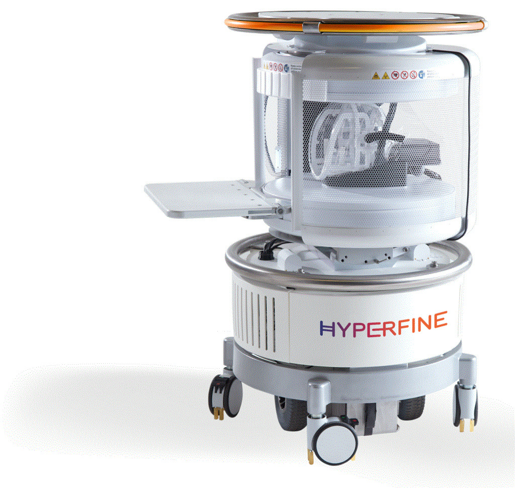
 Hyperfine gets FDA nod for world's first portable bedside MR system
In February, Hyperfine received FDA approval for the world’s first bedside MR system, creating new possibilities for hospitals seeking point-of-care imaging with a system the company says is 20 times cheaper, 10 times lighter and consumes 35 times less power than fixed conventional MR solutions.
The system, which is equipped with a .064 Tesla magnet, requires no shielding. It was developed with the nearly 90% of the world lacking access to MR in mind, but may also prove beneficial for a variety of patients receiving care in places where conventional MR is already available.
"In hospitals, the first applications most people think of is pediatrics and avoiding the ionizing radiation of CT," Chris Ward, head of marketing for Hyperfine, told HCB News. "Another is intensive care where MR would be used if only it were readily accessible for some of the sickest, most fragile and hard-to-transport patients in the hospital."
Prior to its approval by the FDA, thousands of scans were performed as part of investigational partnerships at Yale New Haven University, Penn Medicine, Good Samaritan Hospital Long Island, New York Presbyterian Brooklyn Methodist Hospital, and Brown University.
In September, a team at Yale New Haven published research showing the scanner found evidence of ischemic stroke, hemorrhagic stroke, subarachnoid hemorrhage, traumatic brain injury, and brain tumors in patients who showed signs of neurological symptoms. It also was used to analyze 20 patients with severe COVID-19 symptoms, many of whom were too sick to be moved to an MR suite for a neurological diagnosis. Eight were found to have neurological abnormalities.
Hyperfine unveiled the system at the 2019 RSNA annual meeting.
 Abbreviated MR outperforms 3D mammo dense breast tissue screening
Abbreviated breast MR may be better at detecting cancer in women with dense breast tissue than digital breast tomosynthesis, according to a study published in the Journal of the American Medical Association in late February.
The assessment was made from the findings of 48 centers in the U.S. and Germany who took part in what is described as the first prospective multicenter study of AB-MR. Leading the study was the Memorial Sloan Kettering Cancer Center, in collaboration with an international research team and the ECOG-ACRIN Cancer Research Group.
“These types of studies are needed when evaluating new tests for adoption and to also inform patients of the risk and benefits when considering whether to have the test,” Dr. Christopher E. Comstock, of MSK and the study’s lead author, told HCB News. “Our study confirmed that AB-MR in women at average risk with dense breast tissue outperforms 3D mammography by a factor of 2.4 times in detecting women with breast cancer. And not only does it find more cancer, but it finds invasive and high-grade cancers not found on the 3D mammography.”
A total of 1,444 women between 40 and 75 enrolled in the trial. All had dense breasts on their prior mammograms and did not currently have breast cancer or any clinical symptoms, such as BRCA status or a family history of breast cancer. Twenty-three women were diagnosed with breast cancer during the first year, with AB-MR diagnosing cancer in 22 out of 23. 3D mammography detected only nine cases, while AB-MR identified all of the cancers found with 3D mammograms except for one early-stage case.
The findings were supported later in the year by research from the Perelman School of Medicine at the University of Pennsylvania, which evaluated 475 asymptomatic women with dense breasts who previously had a negative 3D mammography screening, between 2016 and 2019. In their study, published in the Journal of Clinical Oncology, they reported 13 cancers were detected.
Brain abnormalities found on MR scans of almost half of COVID-19 patients in ICU
A study published in the journal Radiology in May found that close to half of COVID-19 patients admitted to ICUs in Turkey showed signs of brain abnormalities on MR scans.
The discovery was made by neuroradiologists at Istanbul University-Cerrahpasa, who assert that the findings validated symptoms already identified for those with the disease, particularly symptoms related to the nervous system.
“Recent evidence highlights a relatively high percentage (36%) of central nervous system symptoms including headache, altered mental status, acute cerebrovascular disease and epilepsy in patients with COVID-19,” said the authors in their report. “The rate of neurological symptoms is higher in patients with more severe respiratory disease status. The relatively high percentage of neurologic symptoms is concordant with studies showing neurotropism of coronavirus.”
The purpose of the study was to evaluate the neurological condition of COVID-19 patients, as current literature on the subject is limited. Six percent of patients assessed experienced acute stroke, while another 15% had an altered state of mind. The authors believe one factor responsible is the cytokine storm, in which the body attacks its own cells and tissues in addition to fighting off the virus.
The researchers performed MR exams on 235 COVID-19 patients in the ICUs of two academic and six non-academic-affiliated hospitals. Of them, 50 (21%) developed neurological symptoms.
All abnormalities were found in different areas of the brain, including the frontal lobe, parietal lobe, occipital lobe, temporal lobe, insular cortex and cingulate gyrus. Diagnoses that included autoimmune encephalitis, seizure, hypoglycemia and hypoxia were the main differentiator of the group, as it can be accompanied by cortical microhemorrhages and blood-brain barrier breakdown. The findings highlight that COVID-19 may result in infectious or autoimmune encephalitis due to its neurotropic potential.
The authors claim that the study may help increase awareness of a possible relationship between SARS-CoV-2 and neurological abnormalities in infected patients, especially those in the ICU. They warn, however, that comorbid conditions must be accounted for as confounding factors in the neurological abnormalities found, including diabetes mellitus, prolonged ICU stays, and respiratory distress with hypoxia.
 Canon gets clearance for iterative reconstruction tech with 1.5T MR system
Iterative reconstruction technology is a growing trend in MR imaging. By allowing scanners to do more with less data, they enable faster scans without compromising image quality.
In June the FDA gave Canon the green light to integrate Compressed SPEEDER technology, which reduces scan time by applying iterative reconstruction to reconstruct full-resolution images from under-sampled data into its Vantage Orian 1.5T MR system.
The news followed a March announcement that the technology was approved for use on the Vantage Galan 3T MR system.
“Canon Medical's Compressed SPEEDER can reduce scan times by a factor of 2.5,” Tom Naypaur, senior manager for MR Solutions Marketing of Canon Medical Systems USA, told HCB News. "In certain instances, we've seen overall scan time reduction of up to 60% comparing acquisitions with and without Compressed SPEEDER. Of course, scan times will vary from site-to-site depending on the scan protocols.”
In reducing the time of MR scans, Compressed SPEEDER enables more scans to be conducted in a day and allows providers more time in between each exam to help clean and disinfect scanners. Reducing the timing of scans enables greater patient comfort, which may help reduce movement from patients caused by discomfort and thereby enable higher-quality images.
It also supports image acceleration and can be used to avoid unfolding artifacts sometimes seen with standard parallel imaging, or can achieve higher resolution in 2D Fast Spin Echo acquisitions.
Compressed SPEEDER technology is included in an all-new version of M-Power software which is available for the Vantage Orian 1.5T. The scanner comes standard with Windows 10 and embedded cybersecurity solutions from Canon Medical that provide continuous patches and updates from Microsoft to keep patient data secure. The scanner also has whitelisting functions, which give clinicians access only to applications and processes that have been authorized.
Prostate MR and biopsy quality vary by institution, says study
While prostate MR scans are growing in popularity, researchers at the University of Minnesota Medical School published findings in Radiology showing the quality of cancer detection may be inconsistent from one facility to the next.
“What this does say is that at some institutions the process of prostate MR and subsequent biopsy of detected lesions is less robust than others, which may be due to the biopsy itself and the urologist performing that biopsy, the MR quality, the pathologists, or the radiologist interpreting the study,” Dr. Ben Spilseth, associate professor in the department of Radiology at the University of Minnesota Medical School and U of M site coordinator for the study, told HCB News in June.
Spilseth and colleagues reviewed prostate biopsies of more than 3,400 men who had targets identified on prostate MR and altogether had 5,082 lesions. Patients were spread out across 26 institutions and results were collected from the Prostate Imaging Reporting and Data System (PI-RADS).
Biopsy findings showed 1,698 cancers were greater than or equal to 3+4 (International Society of Urological Pathology grade group ≥2) in 2,082 men, by Gleason Score standards. The estimated PPV was 35% for a PI-RADS score greater than or equal to 3, and 49% for a score greater than or equal to 4. The interquartile ranges of PPV at these same PI-RADS score thresholds were 27% – 44% and 27% – 48%, respectively.
The team concluded that the positive predictive value for such tests vary at high rates among different sites, and say that more research is needed to determine how similar outcomes can be achieved at different facilities.
Spilseth said a new certification process currently being developed by the American College of Radiology could help in this effort. He asserted, however, that the most important component is for the radiologist and urologist to be in regular communication with one another to monitor biopsy pathology and compare it to MR findings, so that they can assess and troubleshoot unexpected cases quickly.
Are SPIONs the future of low-field MR imaging?
Low-field MR systems are entering areas like neurological ICUs to assess critically ill patients who cannot be as easily transported to MR rooms, but the images they produce are weaker than conventional systems.
In June, researchers at Massachusetts General Hospital and the University of Sydney in Australia investigated the potential of SPIONs for such scans. Short for superparamagnetic iron oxide nanoparticles, SPIONs are 3,000 times more magnetic than conventional MR contrast agents, which could make them ideal for low-field scanners.
“There do not exist contrast agents that perform well enough at low magnetic fields,” physicist Matthew Rosen, director of the Low-field MR and Hyperpolarized Media Laboratory at the MGH Martinos Center for Biomedical Imaging told HCB News. “Adding contrast to low magnetic field scanners allows the full gamut of the type of scans that people do with high-field. It's a very standard workflow of an oncologist who is trying to study the progress of some brain cancer. They would say, ‘let’s order these imaging sequences, plus contrast.’ And so the plus contrast part is something you can now do at low field.”
Physicists David Waddington, the lead author of the study, and Zdenka Kuncic, both from the University of Sydney, tested their approach in a trial involving healthy lab rats that were scanned with Rosen’s homemade ultra-low-field MR scanner. Each subject was given an initial scan without the contrast agent and another with it. Images from both scans were compared, with kidneys, liver and other organs in the contrast-enhanced scan glowing more brightly than in the non-contrast one.
“I’ve been reaching out to clinical collaborators here,” said Rosen. “There is a lot of enthusiasm for that. People I have spoken to, mostly in neurology, are very interested in this. Probably something like this will happen sooner rather than later. The people who are doing low field MR, such as folks at Yale and people on Long Island at Northwell Health, are all very excited about boosting the obtainable imaging information from this new imaging modality.”
GE Healthcare launches a 7T MR system
GE Healthcare unveiled its SIGNA 7T MR system at the International Society for Magnetic Resonance in Medicine (ISMRM) virtual meeting in August.
Equipped with ultra-high field magnet technology, the 60-cm bore scanner offers approximately five times more power than most clinical systems and is expected to enhance imaging for neurodegenerative diseases like Alzheimer’s disease and mild traumatic brain injury (TBI), as well as extremities.
“The integration of the new MR platform into the SIGNA 7.0T system has resulted in outstanding image quality,” said Garry Gold, professor of radiology at Stanford University, in a statement. “Stanford has been working with GE at the 7.0T field strength for over 15 years and we expect this new platform to be transformative for the next generation of scientists, researchers and clinicians.”
The system is designed to detect subtle structures that may hold significant information for clinicians and researchers alike.
It incorporates UltraG gradient technology, GE’s most powerful, whole-body gradient coil for enabling ultra-high field imaging speed and resolution. It also has the familiarity of SIGNA Works applications platform, which provides clinicians with access to state-of-the art applications such as deep learning-based platform tools like AIR x brain for automated slice positioning and Silent MR imaging. Another feature is its Precision RF transmit and receive architecture, which improves image quality and research flexibility.
In November, SIGNA received FDA approval.
NYU and Facebook make MR 4x faster without losing quality
Back in 2018, fastMRI, a collaboration between Facebook Artificial Intelligence Research and NYU Langone's Department of Radiology, released a large-scale, landmark MR data set with a plan to eventually power it with AI and make MR scans significantly faster.
In August they published research in the American Journal of Roentgenology showing how the team’s AI-generated MR scans are just as effective as a traditional scan, but with the added advantage of speed. Theoretically that means providers could scan more patients in a day and patients could experience less discomfort.
“This study is an important step toward clinical acceptance and utilization of AI-accelerated MR scans because it demonstrates for the first time that AI-generated images are essentially indistinguishable in appearance from standard clinical MR exams, and are interchangeable in regard to diagnostic accuracy,” said Michael P. Recht, professor of radiology at NYU Langone and lead study author. “This marks an exciting paradigm shift in how we are able to improve the patient experience and create images.”
The open-source project comprised 1.5 million de-identified MR knee images from 10,000 scans, and raw measurement data from 1,600 scans, from which the researchers built a neural network and trained it. This included reconstruction of views missed from underlying image structure, similar to the way people interpolate sensory information.
In the study, musculoskeletal radiologists were asked to evaluate two sets of scan results per patient, including one from a standard MR exam and one from the fastMRI model, and were not told which one was created using AI. The images were examined for meniscal tears, ligament abnormalities, and cartilage defects, and also graded for quality. Both sets delivered the same results and led to the same diagnoses, but the radiologists said they preferred the AI image quality over the traditional images.
 Reducing MR exam no-shows with AI
Radiology departments could reduce the number of no-shows they see for scheduled MR appointments with the help of AI predictive analytics.
At least that’s what researchers at Singapore’s Changi General Hospital found in a study published in September—and they did it using what they described as “only a modest amount of data and basic feature engineering”.
"The ability to track appointment no-shows is not, in itself, useful unless one is able to intervene, which is really where the challenge lies," lead author Dr. Chong Le Roy, senior consultant for the department of diagnostic radiology at Changi General Hospital, told HCB News. "It is only with the recent advent of state-of-the-art machine learning algorithms and ready availability of increased computational power in the last few years that makes it possible to be able to predict and thereby allow us to address such problems with interventions."
Le Roy and his colleagues trained and validated their model using records of 32,957 outpatient MR appointments scheduled between January 2016 and December 2018 from Changi General Hospital’s radiology information system.
Finding an overall no-show rate of 17.4%, the team assessed various machine learning predictive models developed with widely used open-source software tools before finding a decision tree-based ensemble algorithm that used a gradient boosting framework, XGBoost, version 0.8. They then implemented telephone call reminders over a six-month period for patients whom the model predicted were among the top 25% highest risk for not showing up to their MR appointments.
Following those six months, the no-show rate of the predictive model was 15.9%, compared to 19.3% over the previous year, corresponding to a 17.2% improvement from the baseline no-show rate.
"General radiology subspecialties providing high-volume imaging services utilizing advanced imaging modalities such as magnetic resonance imaging, CT and ultrasound scans where there is a large proportion of ambulant outpatients, patients with chronic nonlife-threatening ailments, and routine screening would benefit the most from reducing no-show rates," said Le Roy. "Some examples include musculoskeletal imaging for common internal derangements of various joints, spine imaging for cervical/low back pain and breast screening imaging."
The findings were published in ARRS’ American Journal of Roentgenology.
From ultra-powerful 7 Tesla magnets, to a new generation of portable systems, the frontiers for MR are advancing in both directions. Meanwhile, deep learning and image reconstruction are bringing more insight to every scan. Here, presented in chronological order, are the ten biggest MR stories of the year from our Daily News online.

It isn’t often that medical imaging news crosses over into the mainstream, but that’s what happened when actor Chuck Norris and his wife, Gena O’Kelley, filed a gadolinium-related lawsuit against Bracco Diagnostics Inc., the subsidiary of Bracco Imaging, in 2017. One of the first big stories of 2020 involved their decision to dismiss the suit in January.
The suit, which was filed against 11 drug companies, alleged that injections of the MR contrast agent led Gena to develop Gadolinium Deposition Disease and resulted in numerous hospitalizations, costing the couple $2 million or more in out-of-pocket costs.
Gena alleged the contrast agent led her to develop symptoms that included cognitive impairment, body pain/burning, kidney damage, loss of energy/mobility, and difficulty breathing. She said in 2017 that she continued to require treatment even five years after her last exposure to gadolinium, including controversial and unreimbursed chelation therapy, for the damage she suffered.
Gadolinium long considered completely safe but in 2006 evidence emerged that it could cause nephrogenic systemic fibrosis in patients with renal insufficiency. In 2013, researchers showed the contrast agent could accumulate in the brains of patients undergoing multiple examinations, but experts say there is no evidence yet to suggest harmful effects from retention.
Upon the dismissal, Bracco released a statement expressing its satisfaction over the decision by the Norris family and their attorneys to end litigation. “Bracco takes patient safety very seriously and stands behind the safety of all of its products, including the MR contrast agents ProHance (Gadoteridol) Injection, 279.3 mg/mL and MultiHance.”
No settlement payment was made and each party paid their own costs.
In February, Hyperfine received FDA approval for the world’s first bedside MR system, creating new possibilities for hospitals seeking point-of-care imaging with a system the company says is 20 times cheaper, 10 times lighter and consumes 35 times less power than fixed conventional MR solutions.
The system, which is equipped with a .064 Tesla magnet, requires no shielding. It was developed with the nearly 90% of the world lacking access to MR in mind, but may also prove beneficial for a variety of patients receiving care in places where conventional MR is already available.
"In hospitals, the first applications most people think of is pediatrics and avoiding the ionizing radiation of CT," Chris Ward, head of marketing for Hyperfine, told HCB News. "Another is intensive care where MR would be used if only it were readily accessible for some of the sickest, most fragile and hard-to-transport patients in the hospital."
Prior to its approval by the FDA, thousands of scans were performed as part of investigational partnerships at Yale New Haven University, Penn Medicine, Good Samaritan Hospital Long Island, New York Presbyterian Brooklyn Methodist Hospital, and Brown University.
In September, a team at Yale New Haven published research showing the scanner found evidence of ischemic stroke, hemorrhagic stroke, subarachnoid hemorrhage, traumatic brain injury, and brain tumors in patients who showed signs of neurological symptoms. It also was used to analyze 20 patients with severe COVID-19 symptoms, many of whom were too sick to be moved to an MR suite for a neurological diagnosis. Eight were found to have neurological abnormalities.
Hyperfine unveiled the system at the 2019 RSNA annual meeting.

Abbreviated breast MR may be better at detecting cancer in women with dense breast tissue than digital breast tomosynthesis, according to a study published in the Journal of the American Medical Association in late February.
The assessment was made from the findings of 48 centers in the U.S. and Germany who took part in what is described as the first prospective multicenter study of AB-MR. Leading the study was the Memorial Sloan Kettering Cancer Center, in collaboration with an international research team and the ECOG-ACRIN Cancer Research Group.
“These types of studies are needed when evaluating new tests for adoption and to also inform patients of the risk and benefits when considering whether to have the test,” Dr. Christopher E. Comstock, of MSK and the study’s lead author, told HCB News. “Our study confirmed that AB-MR in women at average risk with dense breast tissue outperforms 3D mammography by a factor of 2.4 times in detecting women with breast cancer. And not only does it find more cancer, but it finds invasive and high-grade cancers not found on the 3D mammography.”
A total of 1,444 women between 40 and 75 enrolled in the trial. All had dense breasts on their prior mammograms and did not currently have breast cancer or any clinical symptoms, such as BRCA status or a family history of breast cancer. Twenty-three women were diagnosed with breast cancer during the first year, with AB-MR diagnosing cancer in 22 out of 23. 3D mammography detected only nine cases, while AB-MR identified all of the cancers found with 3D mammograms except for one early-stage case.
The findings were supported later in the year by research from the Perelman School of Medicine at the University of Pennsylvania, which evaluated 475 asymptomatic women with dense breasts who previously had a negative 3D mammography screening, between 2016 and 2019. In their study, published in the Journal of Clinical Oncology, they reported 13 cancers were detected.
Brain abnormalities found on MR scans of almost half of COVID-19 patients in ICU
A study published in the journal Radiology in May found that close to half of COVID-19 patients admitted to ICUs in Turkey showed signs of brain abnormalities on MR scans.
The discovery was made by neuroradiologists at Istanbul University-Cerrahpasa, who assert that the findings validated symptoms already identified for those with the disease, particularly symptoms related to the nervous system.
“Recent evidence highlights a relatively high percentage (36%) of central nervous system symptoms including headache, altered mental status, acute cerebrovascular disease and epilepsy in patients with COVID-19,” said the authors in their report. “The rate of neurological symptoms is higher in patients with more severe respiratory disease status. The relatively high percentage of neurologic symptoms is concordant with studies showing neurotropism of coronavirus.”
The purpose of the study was to evaluate the neurological condition of COVID-19 patients, as current literature on the subject is limited. Six percent of patients assessed experienced acute stroke, while another 15% had an altered state of mind. The authors believe one factor responsible is the cytokine storm, in which the body attacks its own cells and tissues in addition to fighting off the virus.
The researchers performed MR exams on 235 COVID-19 patients in the ICUs of two academic and six non-academic-affiliated hospitals. Of them, 50 (21%) developed neurological symptoms.
All abnormalities were found in different areas of the brain, including the frontal lobe, parietal lobe, occipital lobe, temporal lobe, insular cortex and cingulate gyrus. Diagnoses that included autoimmune encephalitis, seizure, hypoglycemia and hypoxia were the main differentiator of the group, as it can be accompanied by cortical microhemorrhages and blood-brain barrier breakdown. The findings highlight that COVID-19 may result in infectious or autoimmune encephalitis due to its neurotropic potential.
The authors claim that the study may help increase awareness of a possible relationship between SARS-CoV-2 and neurological abnormalities in infected patients, especially those in the ICU. They warn, however, that comorbid conditions must be accounted for as confounding factors in the neurological abnormalities found, including diabetes mellitus, prolonged ICU stays, and respiratory distress with hypoxia.

Iterative reconstruction technology is a growing trend in MR imaging. By allowing scanners to do more with less data, they enable faster scans without compromising image quality.
In June the FDA gave Canon the green light to integrate Compressed SPEEDER technology, which reduces scan time by applying iterative reconstruction to reconstruct full-resolution images from under-sampled data into its Vantage Orian 1.5T MR system.
The news followed a March announcement that the technology was approved for use on the Vantage Galan 3T MR system.
“Canon Medical's Compressed SPEEDER can reduce scan times by a factor of 2.5,” Tom Naypaur, senior manager for MR Solutions Marketing of Canon Medical Systems USA, told HCB News. "In certain instances, we've seen overall scan time reduction of up to 60% comparing acquisitions with and without Compressed SPEEDER. Of course, scan times will vary from site-to-site depending on the scan protocols.”
In reducing the time of MR scans, Compressed SPEEDER enables more scans to be conducted in a day and allows providers more time in between each exam to help clean and disinfect scanners. Reducing the timing of scans enables greater patient comfort, which may help reduce movement from patients caused by discomfort and thereby enable higher-quality images.
It also supports image acceleration and can be used to avoid unfolding artifacts sometimes seen with standard parallel imaging, or can achieve higher resolution in 2D Fast Spin Echo acquisitions.
Compressed SPEEDER technology is included in an all-new version of M-Power software which is available for the Vantage Orian 1.5T. The scanner comes standard with Windows 10 and embedded cybersecurity solutions from Canon Medical that provide continuous patches and updates from Microsoft to keep patient data secure. The scanner also has whitelisting functions, which give clinicians access only to applications and processes that have been authorized.
Prostate MR and biopsy quality vary by institution, says study
While prostate MR scans are growing in popularity, researchers at the University of Minnesota Medical School published findings in Radiology showing the quality of cancer detection may be inconsistent from one facility to the next.
“What this does say is that at some institutions the process of prostate MR and subsequent biopsy of detected lesions is less robust than others, which may be due to the biopsy itself and the urologist performing that biopsy, the MR quality, the pathologists, or the radiologist interpreting the study,” Dr. Ben Spilseth, associate professor in the department of Radiology at the University of Minnesota Medical School and U of M site coordinator for the study, told HCB News in June.
Spilseth and colleagues reviewed prostate biopsies of more than 3,400 men who had targets identified on prostate MR and altogether had 5,082 lesions. Patients were spread out across 26 institutions and results were collected from the Prostate Imaging Reporting and Data System (PI-RADS).
Biopsy findings showed 1,698 cancers were greater than or equal to 3+4 (International Society of Urological Pathology grade group ≥2) in 2,082 men, by Gleason Score standards. The estimated PPV was 35% for a PI-RADS score greater than or equal to 3, and 49% for a score greater than or equal to 4. The interquartile ranges of PPV at these same PI-RADS score thresholds were 27% – 44% and 27% – 48%, respectively.
The team concluded that the positive predictive value for such tests vary at high rates among different sites, and say that more research is needed to determine how similar outcomes can be achieved at different facilities.
Spilseth said a new certification process currently being developed by the American College of Radiology could help in this effort. He asserted, however, that the most important component is for the radiologist and urologist to be in regular communication with one another to monitor biopsy pathology and compare it to MR findings, so that they can assess and troubleshoot unexpected cases quickly.
Are SPIONs the future of low-field MR imaging?
Low-field MR systems are entering areas like neurological ICUs to assess critically ill patients who cannot be as easily transported to MR rooms, but the images they produce are weaker than conventional systems.
In June, researchers at Massachusetts General Hospital and the University of Sydney in Australia investigated the potential of SPIONs for such scans. Short for superparamagnetic iron oxide nanoparticles, SPIONs are 3,000 times more magnetic than conventional MR contrast agents, which could make them ideal for low-field scanners.
“There do not exist contrast agents that perform well enough at low magnetic fields,” physicist Matthew Rosen, director of the Low-field MR and Hyperpolarized Media Laboratory at the MGH Martinos Center for Biomedical Imaging told HCB News. “Adding contrast to low magnetic field scanners allows the full gamut of the type of scans that people do with high-field. It's a very standard workflow of an oncologist who is trying to study the progress of some brain cancer. They would say, ‘let’s order these imaging sequences, plus contrast.’ And so the plus contrast part is something you can now do at low field.”
Physicists David Waddington, the lead author of the study, and Zdenka Kuncic, both from the University of Sydney, tested their approach in a trial involving healthy lab rats that were scanned with Rosen’s homemade ultra-low-field MR scanner. Each subject was given an initial scan without the contrast agent and another with it. Images from both scans were compared, with kidneys, liver and other organs in the contrast-enhanced scan glowing more brightly than in the non-contrast one.
“I’ve been reaching out to clinical collaborators here,” said Rosen. “There is a lot of enthusiasm for that. People I have spoken to, mostly in neurology, are very interested in this. Probably something like this will happen sooner rather than later. The people who are doing low field MR, such as folks at Yale and people on Long Island at Northwell Health, are all very excited about boosting the obtainable imaging information from this new imaging modality.”
GE Healthcare launches a 7T MR system
GE Healthcare unveiled its SIGNA 7T MR system at the International Society for Magnetic Resonance in Medicine (ISMRM) virtual meeting in August.
Equipped with ultra-high field magnet technology, the 60-cm bore scanner offers approximately five times more power than most clinical systems and is expected to enhance imaging for neurodegenerative diseases like Alzheimer’s disease and mild traumatic brain injury (TBI), as well as extremities.
“The integration of the new MR platform into the SIGNA 7.0T system has resulted in outstanding image quality,” said Garry Gold, professor of radiology at Stanford University, in a statement. “Stanford has been working with GE at the 7.0T field strength for over 15 years and we expect this new platform to be transformative for the next generation of scientists, researchers and clinicians.”
The system is designed to detect subtle structures that may hold significant information for clinicians and researchers alike.
It incorporates UltraG gradient technology, GE’s most powerful, whole-body gradient coil for enabling ultra-high field imaging speed and resolution. It also has the familiarity of SIGNA Works applications platform, which provides clinicians with access to state-of-the art applications such as deep learning-based platform tools like AIR x brain for automated slice positioning and Silent MR imaging. Another feature is its Precision RF transmit and receive architecture, which improves image quality and research flexibility.
In November, SIGNA received FDA approval.
NYU and Facebook make MR 4x faster without losing quality
Back in 2018, fastMRI, a collaboration between Facebook Artificial Intelligence Research and NYU Langone's Department of Radiology, released a large-scale, landmark MR data set with a plan to eventually power it with AI and make MR scans significantly faster.
In August they published research in the American Journal of Roentgenology showing how the team’s AI-generated MR scans are just as effective as a traditional scan, but with the added advantage of speed. Theoretically that means providers could scan more patients in a day and patients could experience less discomfort.
“This study is an important step toward clinical acceptance and utilization of AI-accelerated MR scans because it demonstrates for the first time that AI-generated images are essentially indistinguishable in appearance from standard clinical MR exams, and are interchangeable in regard to diagnostic accuracy,” said Michael P. Recht, professor of radiology at NYU Langone and lead study author. “This marks an exciting paradigm shift in how we are able to improve the patient experience and create images.”
The open-source project comprised 1.5 million de-identified MR knee images from 10,000 scans, and raw measurement data from 1,600 scans, from which the researchers built a neural network and trained it. This included reconstruction of views missed from underlying image structure, similar to the way people interpolate sensory information.
In the study, musculoskeletal radiologists were asked to evaluate two sets of scan results per patient, including one from a standard MR exam and one from the fastMRI model, and were not told which one was created using AI. The images were examined for meniscal tears, ligament abnormalities, and cartilage defects, and also graded for quality. Both sets delivered the same results and led to the same diagnoses, but the radiologists said they preferred the AI image quality over the traditional images.

Radiology departments could reduce the number of no-shows they see for scheduled MR appointments with the help of AI predictive analytics.
At least that’s what researchers at Singapore’s Changi General Hospital found in a study published in September—and they did it using what they described as “only a modest amount of data and basic feature engineering”.
"The ability to track appointment no-shows is not, in itself, useful unless one is able to intervene, which is really where the challenge lies," lead author Dr. Chong Le Roy, senior consultant for the department of diagnostic radiology at Changi General Hospital, told HCB News. "It is only with the recent advent of state-of-the-art machine learning algorithms and ready availability of increased computational power in the last few years that makes it possible to be able to predict and thereby allow us to address such problems with interventions."
Le Roy and his colleagues trained and validated their model using records of 32,957 outpatient MR appointments scheduled between January 2016 and December 2018 from Changi General Hospital’s radiology information system.
Finding an overall no-show rate of 17.4%, the team assessed various machine learning predictive models developed with widely used open-source software tools before finding a decision tree-based ensemble algorithm that used a gradient boosting framework, XGBoost, version 0.8. They then implemented telephone call reminders over a six-month period for patients whom the model predicted were among the top 25% highest risk for not showing up to their MR appointments.
Following those six months, the no-show rate of the predictive model was 15.9%, compared to 19.3% over the previous year, corresponding to a 17.2% improvement from the baseline no-show rate.
"General radiology subspecialties providing high-volume imaging services utilizing advanced imaging modalities such as magnetic resonance imaging, CT and ultrasound scans where there is a large proportion of ambulant outpatients, patients with chronic nonlife-threatening ailments, and routine screening would benefit the most from reducing no-show rates," said Le Roy. "Some examples include musculoskeletal imaging for common internal derangements of various joints, spine imaging for cervical/low back pain and breast screening imaging."
The findings were published in ARRS’ American Journal of Roentgenology.