
Philips BrightView XCT
Orthopedic Update
July 08, 2009
by Kathy Mahdoubi, Senior Correspondent
This report originally appeared in the June 2009 Nuclear Medicine issue of DOTmed Business News
Some might say the status quo of orthopedic imaging remained relatively undisturbed for the better part of the 1980s, and perhaps even the '90s, but in a matter of just a few years, a relatively new hybrid of molecular imaging and computed tomography, SPECT/CT, has emerged and quickly become the new gold standard for bone imaging and the pathology of skeletal disease.
The combination of single photon emission computed tomography (SPECT), and X-ray computed tomography (CT) has been championed by cardiologists since its inception, but the modality has also gained a great deal of ground in orthopedic imaging and bone pathology. SPECT is a highly sensitive technology that captures functional changes associated with a wide variety of bone and joint tissue abnormalities, including inflammation, infection, benign and malignant lesions, and bone metastases. SPECT is also considered a significantly more affordable alternative to PET imaging.
Standalone modalities have limitations
"The problem with general X-ray is that it's not volumetric, and X-ray and CT can't pick up inflammation or infection," says Dominic Smith, Vice President of Nuclear Medicine Global Marketing for Philips Healthcare. "That's why the combination of SPECT and CT is very good for orthopedics."
Magnetic resonance technology is also problematic when applied to orthopedic procedures, particularly if patients have undergone hip or knee replacement or arthroscopic surgery and have metal implants or prosthetics in place, says Smith.
Upon first examination, a practicing physician treating a patient complaining of unexplained bone pain may decide to order an X-ray, "but when a patient is having bone pain and nothing appears in radiographic imaging, the next step is a bone scan," says Dr. Richard Myers, Chairman of the nuclear medicine department at Radiological Associates of Sacramento, a CA-based radiology practice specializing in diagnostic imaging, nuclear medicine and radiation oncology.
SPECT systems are scintigraphic, meaning they employ gamma camera technology to detect gamma rays emitted by injected radioisotopes localized at areas of increased bone metabolism. Unlike planar gamma camera imaging, SPECT uses multi-planar reconstruction algorithms to yield datasets that can formulate 3-D images of bone and joint tissue.
Southeast Nuclear Electronics, an independent service organization just outside of Atlanta, GA, specializes in the installation, service, repair and deinstallation of Siemens, Philips and GE gamma cameras. Paul Eaton, President of Southeast Nuclear Electronics, has been watching SPECT technology develop since the early 1970s.
"The resolution that these cameras are now obtaining has made a real difference in being able to move quickly to spot abnormality," says Eaton.
Advancements in gamma detectors, collimators, computed tomography hardware and software and improvements in SPECT gantry have driven the technology to its current level of optimization, says Eaton.
But standalone SPECT is not without its own caveat. The spatial resolution of SPECT is typically not that high. In addition, bone scans rely on the proper distribution and bone-labeling of radiopharmaceuticals, and can also be affected by photon attenuation and motion artifacts associated with breathing and patient movement, which can further reduce image clarity. When lesions go undefined on a SPECT bone scan, physicians often have to order follow-up CT scans or MRIs. The SPECT/CT hybrid solution has the ability to provide simultaneous "coregistration" of functional (SPECT) as well as high-resolution anatomical (CT) imaging.
According to a recent retrospective study published in the Journal of Nuclear Medicine, 90% of bone lesions that remained undefined by standalone SPECT were clearly identified after conducting a SPECT/CT scan.
"The general consensus in the nuclear medicine community is that SPECT/CT is preliminarily suggested to be superior to SPECT alone in orthopedic imaging," states Myers.
The superiority of SPECT/CT imaging to SPECT alone has been documented since the hybrid technology's earliest clinical trials and the release of the first generation commercial SPECT/CT scanner, GE Healthcare's Infinia Hawkeye, released in 1999. Since then, other manufacturers like Siemens and Philips have joined GE in providing the lion's share of SPECT/CT production.
"SPECT/CT naturally has changed the way we look at orthopedic imaging," said Dr. Partha Ghosh, Molecular Imaging Director of Clinical Marketing for Siemens Healthcare, which offers the Symbia series of SPECT/CT solutions. "Initially with SPECT and now with the addition of diagnostic CT and image fusion, we are able to do comprehensive imaging in one stroke. It improves our diagnostic confidence and is a definite advantage in orthopedic imaging."
Taking SPECT/CT and bone imaging to the next level
Philips Healthcare will be exhibiting the next generation of SPECT/CT at this year's Society of Nuclear Medicine annual meeting in Toronto. Philips' BrightView XCT is an advanced flat-panel CT and SPECT system, capable of detecting the finest hairline fractures at a spatial resolution that far exceeds the current capabilities of SPECT, which are estimated to run between 2 and 4 mm. The CT component of the BrightView, in comparison, offers a spatial resolution of .33 mm, as well as advanced attenuation correction, procedure software especially designed for orthopedics, and an extra-large, rotating gantry.
"Orthopedic surgeons, depending upon the institution, are one of the most powerful groups in the hospital," says Smith. "They're big revenue generators. Orthopedic surgery is a booming area for a lot of institutions and this is the first advancement in orthopedic imaging to come out in a long time."
The BrightView XCT has been in clinical testing for the past six months at the Radiological Associates of Sacramento, where Myers is the principal researcher. Philips is preparing to ship 50 more units of the BrightView XCT throughout the U.S. and internationally by the second half of 2009.
SPECT/CT - a financial watershed
Not only is SPECT/CT and its associated radiopharmaceuticals significantly more affordable than PET, they are also well-reimbursed by the Centers of Medicare and Medicaid Services. New developments in PET/CT reimbursement may begin changing the game for SPECT/CT, but for now SPECT/CT is enjoying much higher rates of clinical adoption and CMS approval.
"[SPECT/CT] is growing very quickly, not only in the U.S., but globally," says Sergio Calvo, Director of Siemens' Molecular Imaging Product Marketing. Calvo estimates that the rate of clinical adoption of SPECT/CT in many developed countries to be about 50%, and the widespread success of the modality has developed over just the past two to three years, he says.
Bone scans in general account for 15% to 20% of procedure volume in the area of nuclear medicine, depending upon the practice, says Smith. That percentage is often higher in hospital imaging departments.
"It has a lot to do with an aging population," explains Smith. "Between extreme sports and people getting a little bit older, ortho procedures are forecast to be increasing. We're also seeing a lot of reprocedures because of advancements in technology in hip and knee replacement."
The bone-seeking agents of SPECT
The SPECT side of the technology is useless without the phosphate-compound and gamma-emitting radiopharmaceuticals chemically designed to be taken up by bone. SPECT scanning is highly interventional because it not only aids in bone pathology, but also forecasts possible paths of disease and appropriate treatment.
"Functional changes on the bone's surface predispose structural changes. These changes may take weeks and even months to appear," says Dr. Jeffrey P. Norenberg, Executive Director of the National Association of Nuclear Pharmacies and Associate Director of the New Mexico Center for Isotopes in Medicine at the University of New Mexico, Albuquerque.
SPECT/CT enables precise imaging of bone metastases and infection before either has a chance to enter more advanced, destructive stages. SPECT scanning can also guide biopsy procedures and orthopedic surgery and provide a vehicle for administering bone cancer radiotherapy treatments, says Norenberg.
The radiopharmaceuticals used in SPECT bone scans are comprised of the radioisotope technetium-99m and one of two phosphate compounds, methylene diphosphonate (MDP) and hydroxymethylene diphosphonate (HDP). Their generic drug names are medronate and oxidronate, says Dr. Richard Green, Director of Radiopharmacy Practice for the central-Ohio based Cardinal Health, a leading supplier of radiopharmaceuticals.
In clinical studies, both have been shown to be equally effective in bone scintigraphy and are used "almost interchangeably," says Norenberg. "The vast majority of bone surveys, unless there is an interruption in supply, are conducted using MDP or HDP."
Technically, there is currently a shortage of technetium-99m's parent isotope, molybdenum, but the centralized nuclear pharmacy system in the U.S. is able to support the majority of SPECT procedure volume, says Green.
"I would like to have more molybdenum, but we are able to supply about 85% to 90% of our customers' demands," he says.
Once injected with the radioisotope, patients undergoing a SPECT or SPECT/CT procedure can be scanned at multiple stages of agent uptake, often from 30 minutes to 24 hours later, which helps physicians determine specific disease states.
In a typical bone scan there are "hot spots" - areas where there is very high uptake of the agent, which indicates increased bone formation, or osteoblastic activity, and "cold spots," where bone breakdown, or osteolytic lesions, appears to have no labeling whatsoever, says Green.
Orthopedic oncology procedures account for about half of the bone scans conducted at Myers' practice. In certain forms of bone cancer, osteomyeloma, for example, aggressive metastases can lead to necrotic lesions, represented by a cold or dim spot on the scan. In the case of osteosarcoma, a tumor or area of metastases would show-up as an intense hot spot, says Myers.
Infection of the bone, or osteomyelitis, is "an interesting entity," Myers says. At its onset, infections often run cold on a scan, but at some point bone remodeling goes into overdrive and turns red hot. Another radioisotope that can be used with SPECT/CT to scan specifically for infection is indium-111, which homes in on infected bone tissues after being combined with white blood cells, says Green.
The future of SPECT/CT
Perhaps one of the most exciting applications of modern SPECT/CT technology is in the realm of radiotherapy, says Green. Quadramet, another bone-targeting radiopharmaceutical, also known as samarium-153-lexidronam, is the therapeutic version of technetium-99m MDP. Quadramet is a mixed beta-gamma emitter, which means that it has both therapeutic and diagnostic properties and can be used in bone pain palliation procedures, which have been shown to effectively treat painful metastatic bone disease.
In any case, it looks as though any improvements in SPECT/CT will represent a lucky break for an aging population.
Some might say the status quo of orthopedic imaging remained relatively undisturbed for the better part of the 1980s, and perhaps even the '90s, but in a matter of just a few years, a relatively new hybrid of molecular imaging and computed tomography, SPECT/CT, has emerged and quickly become the new gold standard for bone imaging and the pathology of skeletal disease.
The combination of single photon emission computed tomography (SPECT), and X-ray computed tomography (CT) has been championed by cardiologists since its inception, but the modality has also gained a great deal of ground in orthopedic imaging and bone pathology. SPECT is a highly sensitive technology that captures functional changes associated with a wide variety of bone and joint tissue abnormalities, including inflammation, infection, benign and malignant lesions, and bone metastases. SPECT is also considered a significantly more affordable alternative to PET imaging.
Standalone modalities have limitations
"The problem with general X-ray is that it's not volumetric, and X-ray and CT can't pick up inflammation or infection," says Dominic Smith, Vice President of Nuclear Medicine Global Marketing for Philips Healthcare. "That's why the combination of SPECT and CT is very good for orthopedics."
Magnetic resonance technology is also problematic when applied to orthopedic procedures, particularly if patients have undergone hip or knee replacement or arthroscopic surgery and have metal implants or prosthetics in place, says Smith.
Upon first examination, a practicing physician treating a patient complaining of unexplained bone pain may decide to order an X-ray, "but when a patient is having bone pain and nothing appears in radiographic imaging, the next step is a bone scan," says Dr. Richard Myers, Chairman of the nuclear medicine department at Radiological Associates of Sacramento, a CA-based radiology practice specializing in diagnostic imaging, nuclear medicine and radiation oncology.
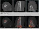
Osteosarcoma of the knee.
0.33 mm thick isotropic
CT slices acquired from
a Philips Brightview XCT
0.33 mm thick isotropic
CT slices acquired from
a Philips Brightview XCT
SPECT systems are scintigraphic, meaning they employ gamma camera technology to detect gamma rays emitted by injected radioisotopes localized at areas of increased bone metabolism. Unlike planar gamma camera imaging, SPECT uses multi-planar reconstruction algorithms to yield datasets that can formulate 3-D images of bone and joint tissue.
Southeast Nuclear Electronics, an independent service organization just outside of Atlanta, GA, specializes in the installation, service, repair and deinstallation of Siemens, Philips and GE gamma cameras. Paul Eaton, President of Southeast Nuclear Electronics, has been watching SPECT technology develop since the early 1970s.
"The resolution that these cameras are now obtaining has made a real difference in being able to move quickly to spot abnormality," says Eaton.
Advancements in gamma detectors, collimators, computed tomography hardware and software and improvements in SPECT gantry have driven the technology to its current level of optimization, says Eaton.
But standalone SPECT is not without its own caveat. The spatial resolution of SPECT is typically not that high. In addition, bone scans rely on the proper distribution and bone-labeling of radiopharmaceuticals, and can also be affected by photon attenuation and motion artifacts associated with breathing and patient movement, which can further reduce image clarity. When lesions go undefined on a SPECT bone scan, physicians often have to order follow-up CT scans or MRIs. The SPECT/CT hybrid solution has the ability to provide simultaneous "coregistration" of functional (SPECT) as well as high-resolution anatomical (CT) imaging.
According to a recent retrospective study published in the Journal of Nuclear Medicine, 90% of bone lesions that remained undefined by standalone SPECT were clearly identified after conducting a SPECT/CT scan.
"The general consensus in the nuclear medicine community is that SPECT/CT is preliminarily suggested to be superior to SPECT alone in orthopedic imaging," states Myers.
The superiority of SPECT/CT imaging to SPECT alone has been documented since the hybrid technology's earliest clinical trials and the release of the first generation commercial SPECT/CT scanner, GE Healthcare's Infinia Hawkeye, released in 1999. Since then, other manufacturers like Siemens and Philips have joined GE in providing the lion's share of SPECT/CT production.
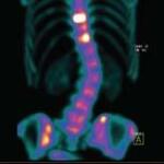
Bone scan acquired from
a Siemens Symbia SPECT/CT
a Siemens Symbia SPECT/CT
"SPECT/CT naturally has changed the way we look at orthopedic imaging," said Dr. Partha Ghosh, Molecular Imaging Director of Clinical Marketing for Siemens Healthcare, which offers the Symbia series of SPECT/CT solutions. "Initially with SPECT and now with the addition of diagnostic CT and image fusion, we are able to do comprehensive imaging in one stroke. It improves our diagnostic confidence and is a definite advantage in orthopedic imaging."
Taking SPECT/CT and bone imaging to the next level
Philips Healthcare will be exhibiting the next generation of SPECT/CT at this year's Society of Nuclear Medicine annual meeting in Toronto. Philips' BrightView XCT is an advanced flat-panel CT and SPECT system, capable of detecting the finest hairline fractures at a spatial resolution that far exceeds the current capabilities of SPECT, which are estimated to run between 2 and 4 mm. The CT component of the BrightView, in comparison, offers a spatial resolution of .33 mm, as well as advanced attenuation correction, procedure software especially designed for orthopedics, and an extra-large, rotating gantry.
"Orthopedic surgeons, depending upon the institution, are one of the most powerful groups in the hospital," says Smith. "They're big revenue generators. Orthopedic surgery is a booming area for a lot of institutions and this is the first advancement in orthopedic imaging to come out in a long time."
The BrightView XCT has been in clinical testing for the past six months at the Radiological Associates of Sacramento, where Myers is the principal researcher. Philips is preparing to ship 50 more units of the BrightView XCT throughout the U.S. and internationally by the second half of 2009.
SPECT/CT - a financial watershed
Not only is SPECT/CT and its associated radiopharmaceuticals significantly more affordable than PET, they are also well-reimbursed by the Centers of Medicare and Medicaid Services. New developments in PET/CT reimbursement may begin changing the game for SPECT/CT, but for now SPECT/CT is enjoying much higher rates of clinical adoption and CMS approval.
"[SPECT/CT] is growing very quickly, not only in the U.S., but globally," says Sergio Calvo, Director of Siemens' Molecular Imaging Product Marketing. Calvo estimates that the rate of clinical adoption of SPECT/CT in many developed countries to be about 50%, and the widespread success of the modality has developed over just the past two to three years, he says.
Bone scans in general account for 15% to 20% of procedure volume in the area of nuclear medicine, depending upon the practice, says Smith. That percentage is often higher in hospital imaging departments.
"It has a lot to do with an aging population," explains Smith. "Between extreme sports and people getting a little bit older, ortho procedures are forecast to be increasing. We're also seeing a lot of reprocedures because of advancements in technology in hip and knee replacement."
The bone-seeking agents of SPECT
The SPECT side of the technology is useless without the phosphate-compound and gamma-emitting radiopharmaceuticals chemically designed to be taken up by bone. SPECT scanning is highly interventional because it not only aids in bone pathology, but also forecasts possible paths of disease and appropriate treatment.
"Functional changes on the bone's surface predispose structural changes. These changes may take weeks and even months to appear," says Dr. Jeffrey P. Norenberg, Executive Director of the National Association of Nuclear Pharmacies and Associate Director of the New Mexico Center for Isotopes in Medicine at the University of New Mexico, Albuquerque.
SPECT/CT enables precise imaging of bone metastases and infection before either has a chance to enter more advanced, destructive stages. SPECT scanning can also guide biopsy procedures and orthopedic surgery and provide a vehicle for administering bone cancer radiotherapy treatments, says Norenberg.
The radiopharmaceuticals used in SPECT bone scans are comprised of the radioisotope technetium-99m and one of two phosphate compounds, methylene diphosphonate (MDP) and hydroxymethylene diphosphonate (HDP). Their generic drug names are medronate and oxidronate, says Dr. Richard Green, Director of Radiopharmacy Practice for the central-Ohio based Cardinal Health, a leading supplier of radiopharmaceuticals.
In clinical studies, both have been shown to be equally effective in bone scintigraphy and are used "almost interchangeably," says Norenberg. "The vast majority of bone surveys, unless there is an interruption in supply, are conducted using MDP or HDP."
Technically, there is currently a shortage of technetium-99m's parent isotope, molybdenum, but the centralized nuclear pharmacy system in the U.S. is able to support the majority of SPECT procedure volume, says Green.
"I would like to have more molybdenum, but we are able to supply about 85% to 90% of our customers' demands," he says.
Once injected with the radioisotope, patients undergoing a SPECT or SPECT/CT procedure can be scanned at multiple stages of agent uptake, often from 30 minutes to 24 hours later, which helps physicians determine specific disease states.
In a typical bone scan there are "hot spots" - areas where there is very high uptake of the agent, which indicates increased bone formation, or osteoblastic activity, and "cold spots," where bone breakdown, or osteolytic lesions, appears to have no labeling whatsoever, says Green.
Orthopedic oncology procedures account for about half of the bone scans conducted at Myers' practice. In certain forms of bone cancer, osteomyeloma, for example, aggressive metastases can lead to necrotic lesions, represented by a cold or dim spot on the scan. In the case of osteosarcoma, a tumor or area of metastases would show-up as an intense hot spot, says Myers.
Infection of the bone, or osteomyelitis, is "an interesting entity," Myers says. At its onset, infections often run cold on a scan, but at some point bone remodeling goes into overdrive and turns red hot. Another radioisotope that can be used with SPECT/CT to scan specifically for infection is indium-111, which homes in on infected bone tissues after being combined with white blood cells, says Green.
The future of SPECT/CT
Perhaps one of the most exciting applications of modern SPECT/CT technology is in the realm of radiotherapy, says Green. Quadramet, another bone-targeting radiopharmaceutical, also known as samarium-153-lexidronam, is the therapeutic version of technetium-99m MDP. Quadramet is a mixed beta-gamma emitter, which means that it has both therapeutic and diagnostic properties and can be used in bone pain palliation procedures, which have been shown to effectively treat painful metastatic bone disease.
In any case, it looks as though any improvements in SPECT/CT will represent a lucky break for an aging population.