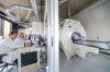
Combined magnetic resonance
imaging (MRI)/operating room
(OR) suite for image-guided
neurosurgery
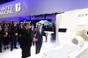
Andover, Mass. and Westchester, Ill. - The Montreal Children's Hospital of the McGill University Health Centre officially opened its new combined magnetic resonance imaging (MRI)/operating room (OR) suite for image-guided neurosurgery. Philips Achieva 3.0T X Series MRI scanner coupled with BrainSUITE® iMRI is the first combination of its kind in North America. The suite was developed in partnership with BrainLAB and Royal Philips Electronics (NYSE: PHG; AEX: PHI) to offer neurosurgeons at The Montreal Children's Hospital a tool to help increase surgical precision in the removal of brain tumors.
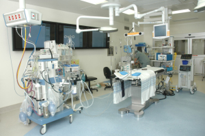
BrainSUITE iMRI provides a real-time "road map" for surgeons in the OR by integrating intra-operative imaging with surgical navigation, planning and digital data management in the OR.
"The powerful combination of BrainSUITE iMRI and the Philips Achieva scanner offers The Montreal Children's Hospital clinically-advanced technology to continue to provide quality care to young patients," stated Sean Clark, president, BrainLAB Inc.


Ad Statistics
Times Displayed: 47765
Times Visited: 1458 MIT labs, experts in Multi-Vendor component level repair of: MRI Coils, RF amplifiers, Gradient Amplifiers Contrast Media Injectors. System repairs, sub-assembly repairs, component level repairs, refurbish/calibrate. info@mitlabsusa.com/+1 (305) 470-8013
BrainSUITE iMRI uses a digital navigation system to show the spatial position of surgical instruments combined with the 3D MRI images of the brain acquired by the scanner. The high-resolution representation of a tumor's location, both prior to and during the surgery, helps guide the removal of tumors.
"Traditionally, during brain surgery, we are guided by MR images taken prior to the procedure. But during surgery, the brain can actually shift as a result of a slight movement of the head, retraction of the brain, or the draining of cerebrospinal fluid. Thus the images the neurosurgeon is relying on are no longer as precise later during the case," said Dr. Jean-Pierre Farmer, Chief of Surgery and a member of the neurosurgery team. "With BrainSUITE iMRI, we have access to images of the brain in real-time which allow us to be much more accurate at determining where the tumor begins and ends. Furthermore the 3 Tesla technology of the new magnet allows us to identify eloquent areas that we need to avoid entering as we resect tumors."
Advanced intra-operative imaging sequences, such as functional MRI (fMRI) and diffusion imaging, also help surgeons identify critical functional areas and plan their approach to a lesion. fMRI depicts what function is located where in the brain and diffusion imaging visualizes brain structures and how different parts of the brain are connected. Providing surgeons with this information helps them remove tumors more efficiently and identify tissue to avoid. The design of Philips' MRI scanner allows for flexibility in patient positioning and access during advanced surgical procedures to further support surgeons in the OR.