
'The fastest growing imaging
equipment segment'
equipment segment'
Top 6 trends impacting CT market growth
January 25, 2017
By Gus Iversen and Tom Dworetzky
The growing number of clinical applications for CT — coupled with an aging population with significant diagnostic needs, and a growing rate of chronic disease — is credited with propelling the global medical CT market to a value of $4.9 billion in 2016.
"Among all medical imaging systems, CT scanners are expected to be the fastest growing imaging equipment segment," said Bruce Carlson, publisher of Kalorama Information, a health care market research source. "Technological developments in the last decade have made it possible to acquire very high quality images in a very short time."
Here are the trends that Carlson and the market analysts at Kalorama Information believe are shaping — or are poised to begin shaping — the CT market.
1. The rise of high slice-count systems
Despite the push to cut health care costs, facilities that have taken the plunge into 256- or 320-slice systems are lauding the superior image quality and saying these deluxe systems lead to better diagnoses in CT angiography, according to the report.
In a perfect value-based world, the improved diagnoses taken from these high powered systems, such as the Aquilion ONE 320 from Toshiba Medical Systems, would be reflected in higher reimbursement.
Still, the report warns that although high slice-count systems are turning lots of heads and generating curiosity, there are still many imaging facilities that wonder if the higher price tag justifies the benefits — particularly when a 64-slice system, for example, can still make a cardiac diagnosis.
Next: Computer software reduces dose
2. Computer software aids in reducing dose
Through initiatives like Image Gently and Image Wisely, limiting lifetime ionizing radiation dose exposure has moved into center stage as a key objective in medical imaging — that means performing exams more efficiently and cutting down on unneeded scans.
Today, there is an entire software market segment dedicated to getting as much as possible from a CT image with radiation doses 'as low as reasonably achievable.' That includes solutions for iterative reconstruction, intraoperativity, and dose-tracking.

With the advent of XR-29, providers who are not incorporating dose monitoring into their CT scanners are being penalized for it. As of January, it can cost them 15 percent of their reimbursement on certain exams.
Last May, HCB News reported that Dr. Ruogu Fang applied machine learning and mathematical algorithms to manipulate low-dose CT perfusion images on stroke patients. When reconstructed, these low-dose images provided comparable image quality with just eight percent of the radiation needed to produce traditional, high-dose images.
There have also been CT studies of late that show that scanning can be of benefit in certain populations to catch cancer earlier, which shows both the risks and rewards of the modality.
Next: Imaging wisely with pediatrics
3. Imaging wisely with pediatrics
From 1996 through the first decade of the new millennium, CT has increasingly been used in pediatric settings, but that trend took a turn, and in recent years the numbers have come down. There is limited evidence regarding the appropriateness of most CT procedures in pediatric patients, according to the report, and as yet it is not clear how much further to restrict its use in this sensitive population.
“Moreover, radiation doses from pediatric CT vary widely in clinical practice,” advised Kalorama, adding that standardization of clinical use and practice of “readily available” dose-reduction strategies could dramatically reduce future radiation-induced cancers caused by CT use in pediatrics.

Like initiatives to promote dose monitoring and iterative reconstruction, carefully selecting pediatric patients for CT imaging is about being cautious and limiting unnecessary exposure.
Last October, a study was presented at the American Academy of Pediatrics National Conference in San Francisco by researchers at the Children’s Hospital of Pittsburgh. They looked at data from 2007 to 2014 and found that while more children were coming to the ED with headaches, fewer CT scans were being done.
Next: Ultra-fast systems
4. Ultra-fast systems
CT is being used more as image quality and speed improve. “Helical scanning and multidetector row CT have led to a tremendous improvement in the speed with which 3D volume can be imaged, and much better routine spatial resolution in the slice direction,” advised the report, adding that in combination with other advances, imaging speed has seen a “phenomenal increase” – essentially exponential – since the early 1970s.
Like the 320 and 640 slice systems, ultra-fast systems might not seem like necessary investments for the average imaging facility, but for those who adopt them, the benefits are clear.

“This increase in speed, along with improvements in low-contrast detectability and image quality, has allowed the technique to be much more robust and this, in turn, has enabled CT to becomemain stream in medical care,” according to the report.
Examples of high speed systems include the Siemens SOMATOM Drive dual source CT scanner and GE Healthcare's Revolution GSI system.
Next: Better resolutions with smart photons
5. Better resolution with smart photons
There are continued improvements that deal with limitations due to "crosstalk" from CT's utlization of reflectors. New direct conversion photon-counting detectors serve as an excellent solution to this issue because the charge carriers produced in the semiconductor follow electric field lines, allowing them to overcome inefficiencies found in scintillator-photodiode detectors used in current commercial systems.
This lets them “readily achieve much better spatial resolution,” according to Kalorama.
Unlike conventional CT scanners, which utilize energy-integrating detectors (EIDs) to create an electrical signal using X-rays, photon-counting detectors calculate individual photon interactions through high-speed semiconductors, eliminating reliance on resolution-limiting scintillator crystals.

The Clinical Center at the National Institutes of Health (NIH) in Maryland, has begun investigating the potential of a photon-counting CT scanner in a hospital-based research setting. It has outlined three areas where photon CT may yield extra diagnostic value:
•Doctors can identify materials in the body with anatomic precision. A dye, or contrast, is often given to a patient so that researchers can see a selected area in more detail. Different materials in the body can be displayed in different colors for faster diagnosis and precision.
•The new technology may be used to help identify and characterize tumors, plaques, or vessels that are smaller than half a millimeter. For many patients, finding a tumor that size may make a difference in identifying if it is benign or could be cancerous.
•The technology may help to more accurately identify soft tissues such as proteins, tendons or collagen which are hard to differentiate with current equipment.
Next: Portable CT units
6. Portable CT units
Kalorama reports that it is “likely” that portable units will become standard in the next 10 years – especially as the speed of diagnosis and treatment becomes more important in improving patient outcomes and cutting health care costs.
Last September, Phil Sullivan, president and CEO of Samsung NeuroLogica, went into detail on the myriad benefits that come with investing in a dedicated portable CT scanner.
These are just a few of the benefits he outlined:

• Patients don’t have to be moved to another part of the building to be scanned for either proton therapy or brachytherapy, saving time and, in some cases, reducing anesthesia.
• With some portable scanners, they don’t have to be moved from one table to another, thus reducing the risk of brachytherapy applicators shifting out of place, and for both proton therapy and brachytherapy patients as well as staff, reducing the physical risks of moving patients.
• Less time for treatment and less moving of patients equates to a more comfortable and efficient treatment experience.
• Specific to brachytherapy, when applicators are out of place and need to be readjusted, in-suite scanners can give closer-to-real-time confirmation that they’ve been correctly repositioned. Patients undergoing cancer treatments have enough worries. Reducing treatment time and the amount of movement are quality- of-care improvements that help prevent adding more anxiety to the process.
To learn more about the CT market, or to purchase the complete report from Kalorama Information, click here
The growing number of clinical applications for CT — coupled with an aging population with significant diagnostic needs, and a growing rate of chronic disease — is credited with propelling the global medical CT market to a value of $4.9 billion in 2016.
"Among all medical imaging systems, CT scanners are expected to be the fastest growing imaging equipment segment," said Bruce Carlson, publisher of Kalorama Information, a health care market research source. "Technological developments in the last decade have made it possible to acquire very high quality images in a very short time."
Here are the trends that Carlson and the market analysts at Kalorama Information believe are shaping — or are poised to begin shaping — the CT market.
1. The rise of high slice-count systems
Despite the push to cut health care costs, facilities that have taken the plunge into 256- or 320-slice systems are lauding the superior image quality and saying these deluxe systems lead to better diagnoses in CT angiography, according to the report.
In a perfect value-based world, the improved diagnoses taken from these high powered systems, such as the Aquilion ONE 320 from Toshiba Medical Systems, would be reflected in higher reimbursement.
Still, the report warns that although high slice-count systems are turning lots of heads and generating curiosity, there are still many imaging facilities that wonder if the higher price tag justifies the benefits — particularly when a 64-slice system, for example, can still make a cardiac diagnosis.
2. Computer software aids in reducing dose
Through initiatives like Image Gently and Image Wisely, limiting lifetime ionizing radiation dose exposure has moved into center stage as a key objective in medical imaging — that means performing exams more efficiently and cutting down on unneeded scans.
Today, there is an entire software market segment dedicated to getting as much as possible from a CT image with radiation doses 'as low as reasonably achievable.' That includes solutions for iterative reconstruction, intraoperativity, and dose-tracking.

Dose knowledge is crucial
With the advent of XR-29, providers who are not incorporating dose monitoring into their CT scanners are being penalized for it. As of January, it can cost them 15 percent of their reimbursement on certain exams.
Last May, HCB News reported that Dr. Ruogu Fang applied machine learning and mathematical algorithms to manipulate low-dose CT perfusion images on stroke patients. When reconstructed, these low-dose images provided comparable image quality with just eight percent of the radiation needed to produce traditional, high-dose images.
There have also been CT studies of late that show that scanning can be of benefit in certain populations to catch cancer earlier, which shows both the risks and rewards of the modality.
3. Imaging wisely with pediatrics
From 1996 through the first decade of the new millennium, CT has increasingly been used in pediatric settings, but that trend took a turn, and in recent years the numbers have come down. There is limited evidence regarding the appropriateness of most CT procedures in pediatric patients, according to the report, and as yet it is not clear how much further to restrict its use in this sensitive population.
“Moreover, radiation doses from pediatric CT vary widely in clinical practice,” advised Kalorama, adding that standardization of clinical use and practice of “readily available” dose-reduction strategies could dramatically reduce future radiation-induced cancers caused by CT use in pediatrics.

Protecting children from
radiation exposure
radiation exposure
Like initiatives to promote dose monitoring and iterative reconstruction, carefully selecting pediatric patients for CT imaging is about being cautious and limiting unnecessary exposure.
Last October, a study was presented at the American Academy of Pediatrics National Conference in San Francisco by researchers at the Children’s Hospital of Pittsburgh. They looked at data from 2007 to 2014 and found that while more children were coming to the ED with headaches, fewer CT scans were being done.
4. Ultra-fast systems
CT is being used more as image quality and speed improve. “Helical scanning and multidetector row CT have led to a tremendous improvement in the speed with which 3D volume can be imaged, and much better routine spatial resolution in the slice direction,” advised the report, adding that in combination with other advances, imaging speed has seen a “phenomenal increase” – essentially exponential – since the early 1970s.
Like the 320 and 640 slice systems, ultra-fast systems might not seem like necessary investments for the average imaging facility, but for those who adopt them, the benefits are clear.
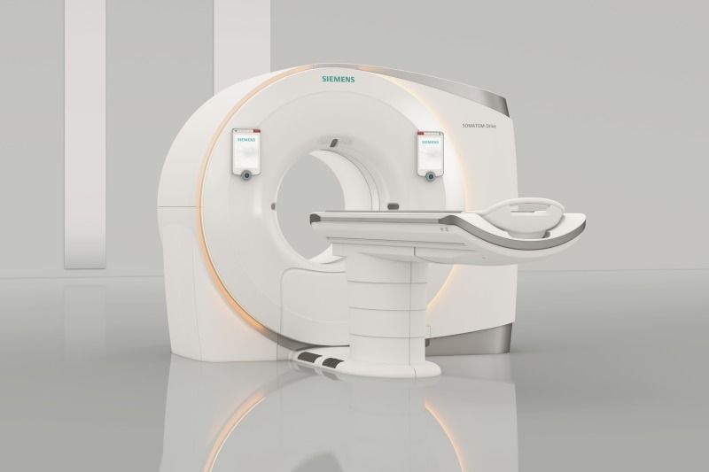
Siemens SOMATOM Drive CT
“This increase in speed, along with improvements in low-contrast detectability and image quality, has allowed the technique to be much more robust and this, in turn, has enabled CT to becomemain stream in medical care,” according to the report.
Examples of high speed systems include the Siemens SOMATOM Drive dual source CT scanner and GE Healthcare's Revolution GSI system.
5. Better resolution with smart photons
There are continued improvements that deal with limitations due to "crosstalk" from CT's utlization of reflectors. New direct conversion photon-counting detectors serve as an excellent solution to this issue because the charge carriers produced in the semiconductor follow electric field lines, allowing them to overcome inefficiencies found in scintillator-photodiode detectors used in current commercial systems.
This lets them “readily achieve much better spatial resolution,” according to Kalorama.
Unlike conventional CT scanners, which utilize energy-integrating detectors (EIDs) to create an electrical signal using X-rays, photon-counting detectors calculate individual photon interactions through high-speed semiconductors, eliminating reliance on resolution-limiting scintillator crystals.
Photon counting CT at NIH
The Clinical Center at the National Institutes of Health (NIH) in Maryland, has begun investigating the potential of a photon-counting CT scanner in a hospital-based research setting. It has outlined three areas where photon CT may yield extra diagnostic value:
•Doctors can identify materials in the body with anatomic precision. A dye, or contrast, is often given to a patient so that researchers can see a selected area in more detail. Different materials in the body can be displayed in different colors for faster diagnosis and precision.
•The new technology may be used to help identify and characterize tumors, plaques, or vessels that are smaller than half a millimeter. For many patients, finding a tumor that size may make a difference in identifying if it is benign or could be cancerous.
•The technology may help to more accurately identify soft tissues such as proteins, tendons or collagen which are hard to differentiate with current equipment.
6. Portable CT units
Kalorama reports that it is “likely” that portable units will become standard in the next 10 years – especially as the speed of diagnosis and treatment becomes more important in improving patient outcomes and cutting health care costs.
Last September, Phil Sullivan, president and CEO of Samsung NeuroLogica, went into detail on the myriad benefits that come with investing in a dedicated portable CT scanner.
These are just a few of the benefits he outlined:
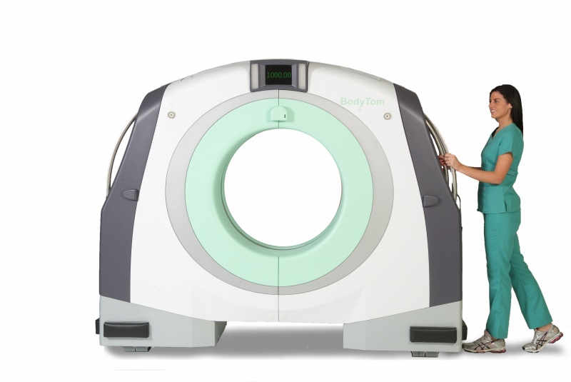
NeuroLogica BodyTom
portable CT
portable CT
• Patients don’t have to be moved to another part of the building to be scanned for either proton therapy or brachytherapy, saving time and, in some cases, reducing anesthesia.
• With some portable scanners, they don’t have to be moved from one table to another, thus reducing the risk of brachytherapy applicators shifting out of place, and for both proton therapy and brachytherapy patients as well as staff, reducing the physical risks of moving patients.
• Less time for treatment and less moving of patients equates to a more comfortable and efficient treatment experience.
• Specific to brachytherapy, when applicators are out of place and need to be readjusted, in-suite scanners can give closer-to-real-time confirmation that they’ve been correctly repositioned. Patients undergoing cancer treatments have enough worries. Reducing treatment time and the amount of movement are quality- of-care improvements that help prevent adding more anxiety to the process.
To learn more about the CT market, or to purchase the complete report from Kalorama Information, click here