Therapeutic Technologies closeout auctions take place on DOTmed.com. Bid today on 538 lots of medical equipment. Inspections may be done by appointment. Hours are Monday through Friday 9am-4pm Contact chrisnuytens1@gmail.com to schedule.

C2 Management processes all types of equipment, from a few pallets of surplus items to large industrial generators. We specialize in maximizing reuse, handling everything from IT equipment to heavy machinery with efficiency & sustainability in mind.

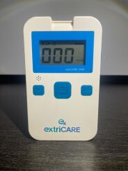
DEVON extriCARE 2400 Negative Pressure Wound Therapy REF: 111512 "The sale of this item may be subject to regulation by the U.S. Food and Drug Administration and state and local regulatory... view more
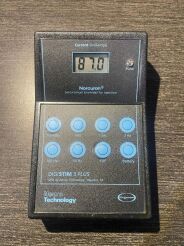
Neuro Technology Tens Unit Digistim 3 Plus. BO‑7200 "The sale of this item may be subject to regulation by the U.S. Food and Drug Administration and state and local regulatory agencies. If so,... view more
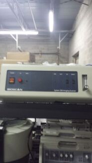
BioMed Certified BioScan System 200 Imaging Scanner in good condition. Turns on but not tested. Selling "As Is" equipment. Contact me with any... view more
Asking Price:
$150 USD

MTS MICRO TYPING SYSTEMS 5150-60 CENTRIFUGE Only parts The unit powers on, but the door does not lock. sold as-is. Important Notice: This item is only available for purchase by licensed... view more
Asking Price:
$110 USD

Add to your cart for a shipping quote. Sold as is/as pictured, no additional parts/cables included. SELLER has not independently tested this unit. Please let us know if you have any questions... view more
Asking Price:
$400 USD

Our products are intended for professional use only Shipping fees are calculated automatically based on your location at checkout. Combined shipping rates Available when you purchase multiple... view more
Asking Price:$625 USD
$369 USD (41% Off)
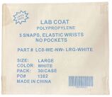
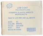
REF LCO-WE-NW- LRG-WHITE LAB COAT POLYPROPYLENE 5 SNAPS, ELASTIC WRISTS NO POCKETS, 30/CASE WHEN EXPIRED FOR TEACHING PURPOSE ONLY NOT FOR HUMAN USE, FOR EDUCATIONAL USE ONLY We entertain... view more
Asking Price:$27 USD
$24 USD (10% Off)
REF LCO-WE-NW-XL-WHITE LAB COAT POLYPROPYLENE 5 SNAPS, ELASTIC WRISTS NOPOCKETS, 30/CASE WHEN EXPIRED FOR TEACHING PURPOSE ONLY NOT FOR HUMAN USE, FOR EDUCATIONAL USE ONLY We entertain all... view more
Asking Price:$27 USD
$24 USD (10% Off)
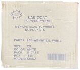
REF LCO-WE-NW-2XL-WHITE LAB COAT POLYPROPYLENE 5 SNAPS, ELASTIC WRISTS NO POCKETS, 30/CASE WHEN EXPIRED FOR TEACHING PURPOSE ONLY NOT FOR HUMAN USE, FOR EDUCATIONAL USE ONLY We entertain... view more
Asking Price:$27 USD
$24 USD (10% Off)

VEBA Meditemp Bagwarmer / VebaBox 3 Units Available Condition: Used – Visible signs of wear / cosmetic damage... view more
Asking Price:
€75 EUR

We acquired the units in a state surplus sale. We don’t know the prior usage history. Units may not be complete and may require repairs. Completely untested. One unit has a missing wheel. ... view more
Asking Price:
$17,550 USD

Storage #H74 Functional Condition: Unit came from a working environment and is tested and working properly. I would recommend preventative maintenance be performed and recertification by... view more
Asking Price:
$200 USD
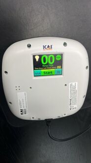
KAI Medical KMS 200 Non-Contact Respiratory RSpot Check 550-0003-01 "The sale of this item may be subject to regulation by the U.S. Food and Drug Administration and state and local... view more
Asking Price:
$300 USD
BAILEY Rib Contractor Large thumbscrew automatic ratchet ( LOT OF 4 ) WHEN EXPIRED FOR TEACHING PURPOSE ONLY NOT FOR HUMAN USE, FOR EDUCATIONAL USE ONLY We entertain all reasonable offers... view more
Asking Price:$295 USD
$174 USD (41% Off)

Smiths Medical Medfusion 3500 Syringe Infusion Pump DOM: 2018-05-10 VER V5.0.0 POST- Pass Buttons, display, indicator lights, audible alarm- Pass Functional test with 60ml B-D syringe @... view more
Asking Price:
$450 USD 


 6
6