by
Lauren Dubinsky, Senior Reporter | August 11, 2015
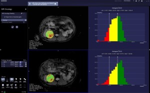
The patient’s histograms
indicate a growth
in area of concern
from the first exam
to the follow-up exam,
indicating that this patient
may not be responding
to therapy
Siemens Healthcare announced yesterday FDA approval of its new MR postprocessing applications — syngo.MR OncoCare for oncology and syngo.MR Neuro Dynamics for neurology.
Oncologists will now be able to use MR to determine a patient’s response to treatment.
“OncoCare’s ability to assess therapy response complements the other inherent strengths of MR — non ionizing radiation exams, multiple contrasts and multi-planar imaging capabilities,” Anne Sheehan, MR product manager for Siemens Medical Solutions, told HCB News. “It’s a powerful new tool for radiologists.”


Ad Statistics
Times Displayed: 46419
Times Visited: 1408 MIT labs, experts in Multi-Vendor component level repair of: MRI Coils, RF amplifiers, Gradient Amplifiers Contrast Media Injectors. System repairs, sub-assembly repairs, component level repairs, refurbish/calibrate. info@mitlabsusa.com/+1 (305) 470-8013
OncoCare is a standalone software package that can be put on a PC or a syngo.via server and it is compatible with all Siemens MR systems. It can be used on every body part ranging from the brain to the liver.
The syngo.MR Neuro Dynamics is an application that provides tools to radiologists to assess tumors in the brain. It can be used to evaluate the blood flows of those tumors.
To use Onco.Care, the radiologist loads the MR studies, draws a region of interest around a tumor and creates histograms using the software, which allow for quantitative evaluation of tumor response. They are then able to follow the patient across a plethora of therapies to determine if they have responded.
MR images show great anatomical detail, but in the past they haven’t been able to follow tumor response. PET imaging is the traditional way in which tumor response is assessed, but it involves radiation.
“You are going to be exposed to imaging that is based on ionizing radiation, but you want to minimize it,” said Sheehan.
MR systems are typically more expensive than PET systems, but this software package can be added on to even the less expensive MR systems.
The software is also helpful for referring physicians because it displays a color circle around the region of interest — red signals an area of concern and green means the area is not of concern. The histograms are for those who want to be involved in the data.