by
John R. Fischer, Senior Reporter | April 27, 2023
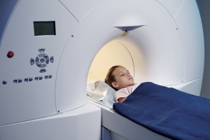
Undergoing four or more CT scans more than doubles a pediatric patient's risk of developing certain cancers.
In a new paper, Taiwanese researchers are reemphasizing the need for ensuring pediatric patients only undergo CT imaging when necessary to avoid overexposing them to radiation, having found that scanning children four or more times more than doubles their risk of developing certain cancers.
Comparing data on 7,807 children under 18 diagnosed with intracranial tumors, leukemia or lymphoma between 2000 and 2013, and 78,057 control subjects, they found that one CT scan had no increased risk of any cancers compared with no exposure, but that two- to three- scans raised the chances of developing intracranial tumors. Those who received four or more had over a two-fold risk of being diagnosed with intracranial tumors, leukemia, and non-Hodgkin's lymphoma.
"Parents and pediatric patients should be well informed on risks and benefits before radiological procedures and encouraged to participate in decision-making around imaging,” wrote Dr. Yu-Hsuan Joni Shao, of the College of Medical Science and Technology at Taipei Medical University, and her coauthors, in their report.


Ad Statistics
Times Displayed: 174492
Times Visited: 3183 For those who need to move fast and expand clinical capabilities -- and would love new equipment -- the uCT 550 Advance offers a new fully configured 80-slice CT in up to 2 weeks with routine maintenance and parts and Software Upgrades for Life™ included.
The authors say that younger children appear to be more at risk of developing cancer from repeated CT scans, and that providers should be careful in their use of this technology and consider using radiation-reducing techniques.
Reducing unnecessary exposure can be achieved through a variety of means, one of which is adopting and abiding by guidelines and standards around appropriate imaging.
The ALARA (As Low As Reasonably Achievable) Principle, for instance, introduced by the International Commission on Radiological Protection in 1977, ensures that patients are exposed to minimum radiation levels during imaging, and spares them from any excess radiation that does not have a direct benefit. It advises that providers minimize time spent performing exams as much as possible, as well as distancing themselves from radioactive sources, and using appropriate shielding technologies and resources to protect themselves and patients, according to the CDC.
But following this risks decreasing the quality of CT images, hindering results. Optimization strategies like creating reference CT images by scanning specifically designed phantoms can help clinicians decipher a balance between radiation exposure and the amount of machine power needed to produce quality CT scans, but tasks like this are often manual and time-consuming.
In March, researchers at the University of Florence and radiologists and medical physicists from Florence Hospital
automated this process with AI, creating a data set of 30,000 labeled CT images captured with different tomographic reconstruction configurations performed on phantoms that mimicked human tissue. They trained two AI models on these configurations and then tested them to see if they assessed CT images as well as humans.