by
John R. Fischer, Senior Reporter | October 17, 2023
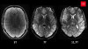
Adjustments in how different MR imaging techniques are performed can improve scan quality and potentially avoid the need for invasive biopsies to confirm prostate cancer diagnoses. (Photo courtesy of University College London)
With a few adjustments in how they capture certain types of MR images, clinicians could help prostate cancer patients avoid invasive and expensive biopsies and save time and costs on scans, according to a new study.
Since 2020, researchers at University College London and University College London Hospital have conducted the PRIME trial to investigate whether a cheaper and less invasive two-step MR scan is as effective as a standard three-phase scan for diagnosing prostate cancer. One part of the trial, the GLIMPSE study has now shown that changes in three MR scanning techniques can improve image quality to the point where clinicians can rule out or identify, and stage cancer without requiring a follow-up biopsy.
Using a five-point scoring system, Prostate Imaging Quality (PI-QUAL), the researchers assessed 355 MR scans from 41 medical centers in 18 countries and found that 32% achieved the highest score of five, indicating optimal quality. Providing feedback, the researchers asked the centers to resubmit their MR scans. Thirty-six complied, with the percentage that scored five rising to 97%, meaning these scans alone could rule out cancer, unlike lower-scored ones.


Ad Statistics
Times Displayed: 174542
Times Visited: 3184 For those who need to move fast and expand clinical capabilities -- and would love new equipment -- the uCT 550 Advance offers a new fully configured 80-slice CT in up to 2 weeks with routine maintenance and parts and Software Upgrades for Life™ included.
Here are the three areas where changes helped raise scores:
- T2-weighted imaging: Adjust in-plane resolution to meet PI-RADS, version 2.1, guidelines; acquire an additional orthogonal separate plane; and decrease section thickness to 3 mm.
- Diffusion-weighted imaging: Acquire or calculate a high b value of more than 1400 sec/mm2; decrease section thickness to 4 mm or less; and reduce the field of view to between 16 and 22 cm.
- Dynamic contrast-enhanced imaging: Adjust temporal resolution to 15 seconds maximum; use fat-suppressed sequences; and use a power injector with a pump speed of 3 mL/second with a saline chaser.
“The quality of MR scans can be significantly improved by following a few simple recommendations. This could be changing the duration of certain sequences by a few seconds, for example,” said first author Dr. Alexander Ng, of the UCL division of medicine, in a statement.
Multiparametric MR scans are the standard for diagnosing prostate cancer and can rule out the need for upfront tissue biopsies, which can be uncomfortable and lead to complications. Should the PRIME study be successful, it is expected to reduce MR scan time from 30 to 20 minutes, on average, as well as reduce clinical staff, make scans less invasive without the need for intravenous contrast, and potentially save on costs for providers.
The PI-QUAL system was developed as part of the UCL-led PRECISION study, a predecessor to the PRIME trial, published in 2018, which led to changes in international clinical practice that allow for all men to receive an upfront MR scan instead of a biopsy.
The findings for GLIMPSE were published in
Radiology.