Nationwide Imaging sells only the finest pre-owned equipment, with C-Arms being our specialty. Your installed system is fine-tuned and worry-free, because we stand behind our work. Get the best for less -- at Nationwide.

Medilab Global provides high-quality pre-owned imaging equipment, parts, and service to the healthcare industry worldwide. Our number 1 position is to provide excellent customer service.. 305-234-0084 | Email: info@medilabglobal.com | Hablamos Español

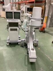
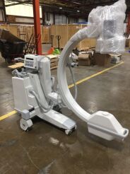
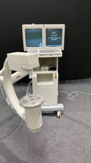
OEC 8800 Flexview C-ARM Sortly ID: SA304T0501 DOM: 2003... view more
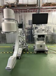
OEC Elite ESP 12”II C-ARM DOM: 2017 Tri Mode Image Intensifier, DICOM 3.0, 1kx1kx16 bit Digital Image Processing, Noise Filter, Real-time Variable DRM Enhancement, Zoom and Roam, Negate Mode, Dual... view more
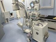
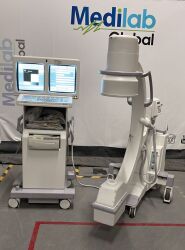
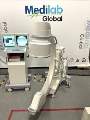
OEC 9900 12”II Elite C-ARM DOM: 2008 Tri Mode Image Intensifier, DICOM 3.0, 1kx1kx16 bit Digital Image Processing, Noise Filter, Real-time Variable DRM Enhancement, Zoom and Roam, Negate Mode, Dual... view more
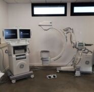
OEC 9900 12”II Elite C-ARM DOM: 2008 System Software: 7.0.31... view more
Over 35,000 parts in stock for all modalities including MRI, CT, Cath Lab, Mammography & C-Arm. Enjoy same day shipping until 7:30PM EST Mon-Fri & 24/7 technical support. Visit https://parts.blockimaging.com to order today! Call Block Imaging 877-621-2887
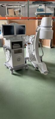
OEC 9600 9”II ESP C-ARM (Fully Refurbished with Flat Screen Monitors) Includes Flat Screen Monitors, Rotating X-ray Tube, Tri-Mode Image Intensifier, High Resolution CCD, Fluoro Boost, Pulsed... view more
OEC Elite ESP 12”II C-ARM DOM: 2017... view more
OEC 8800 FlexiView C-ARM DOM: 2003 ESP Dual Touch Screen Monitors Tri-Mode 9/6/4.5 Inch Image Intensifier Stationary X-Ray Tube 200 Image Storage High Resolution 1k x 1k CCD Camera Dual High... view more

OEC 9600 C-ARM DOM: 1998 Dual Monitors, Tri-Mode 4"/6"/9" Image Intensifier, 200 Image Storage, LIH-Last Image Hold, General Surgical Package, Trace, Fluoro Boost, Sharpen Function, Gamma... view more
Lightly used system available direct from the end user customer. MONARCH QUEST Featuring our most advanced, data-driven navigation software with AI-Powered algorithms & advanced imaging integration with the GE HealthCare OEC 3D C-arm.

OEC 9800 12"II REV 30 C-ARM DOM: 2005 Digital, Dual 1Kx1K Monitors, Right Touch Screen, Tri-Mode, Image Intensifier, 400 Image Storage, LIH-Last Image Hold, Fluoro Boost, Sharpen Function, Gamma... view more

DOM: 2002 Digital, Dual 1Kx1K Monitors, Right Touch Screen, Tri-Mode, Image Intensifier, 400 Image Storage, LIH-Last Image Hold, Fluoro Boost, Sharpen Function, Gamma Correction, Frame Averaging,... view more

OEC 9800 12"II REV 30 C-ARM Digital, Dual 1Kx1K Monitors, Right Touch Screen, Tri-Mode, Image Intensifier, 400 Image Storage, LIH-Last Image Hold, Fluoro Boost, Sharpen Function, Gamma Correction,... view more
DOM: 2003 Digital, Dual 1Kx1K Monitors, Right Touch Screen, Tri-Mode, Image Intensifier, 400 Image Storage, LIH-Last Image Hold, Fluoro Boost, Sharpen Function, Gamma Correction, Frame Averaging,... view more
We provide cost effective solutions for your budget & assist you in buying, selling, site planning, project management, installation, service & maintenance for your C-Arm. For our turn-key solutions call us today 212-366-9100 or info@atlantisworldwide.com

DOM: 1999 Digital, Dual 1Kx1K Monitors, Right Touch Screen, Tri-Mode, Image Intensifier, 400 Image Storage, LIH-Last Image Hold, Fluoro Boost, Sharpen Function, Gamma Correction, Frame Averaging,... view more
DOM: 1996 Dual Monitors, 4” Image Intensifier, 60 Image Storage, LIH-Last Image Hold, Fluoro Boost, Sharpen Function, MARS-Motion Artifact Reduction, Gamma Correction.... view more

DOM: 2008 Digital, Dual 1Kx1K Monitors, Right Touch Screen, Tri-Mode, Image Intensifier, 400 Image Storage, LIH-Last Image Hold, Fluoro Boost, Sharpen Function, Gamma Correction, Frame Averaging,... view more
C-Arm machines are a type of medical imaging device used in orthopedic, vascular, and pain management procedures. It is a type of X-ray machine that is used to take images of the body in a non-invasive manner. The C-Arm is a mobile device that is composed of an X-ray source, an image intensifier, and a digital imaging system. C-Arm machines is used to produce real-time images of the body during a procedure, allowing the physician to make quick decisions and adjustments to the procedure.
C-Arm equipment is used in a variety of medical procedures, including orthopedic surgery, vascular surgery, and pain management. It is also used to diagnose and treat a variety of conditions, including fractures, tumors, and cardiac issues. The C-Arm can also be used to guide minimally invasive procedures, such as catheterization and biopsies.
The price range for C-Arm equipment can vary greatly depending on the type and features of the device. Generally, the cost of a C-Arm can range from $50,000 to $200,000. There are a variety of manufacturers that produce C-Arm equipment, including GE Healthcare, Philips, and Toshiba.


