We are the difference in the market! Real post service. We have our own refurbishment lab with original parts. Specialist in Lasers, Phacos, Microscopes, Slit Lamps, Visual Fields, Retinal Cameras. More than 20 years of experience!. Please Call 3055049163

We have everything you're lookinig for: - refurbished ophthalmmic equipments; - lowest prices in the Market; - immediate and effective contact through whatsapp; - shipping all around the world. +393339997440 commerciale@oftas.it

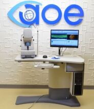
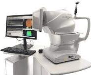
OCT Topcon Triton with Angio, in perfect conditions. Warranty of six months. For more info contact us ... view more
Asking Price:
€22,900 EUR

OCT Topcon 3D 2000, in excellent conditions, with PC and printer. Warranty of six months. For more info contact us ... view more
Asking Price:
€8,500 EUR
topcon 3d 1000 oct power table not included. included : - topcon imagenet software. - hp pc. - lcd led monitor - mouse and keyboard. - dust cover. - guide manual. ... view more
Asking Price:
$19,000 USD
refurbished topcon oct-1 maestro 1 in very good conditions. Warranty 6 months, whipping... view more
Asking Price:
€11,900 EUR 
Oct Topcon Maestro 2 year 2019, in wonderful conditions. For more informations and picture contact us on whatsapp, we can do demo in videocall as well. Warranty 6 months, shipping... view more
Asking Price:
$18,900 USD

Reconditioned (used), Technical condition: very good, Visual condition: very good, Real photos of the product, Made in Japan, (2013), Power supply: 230 V, Frequency: 50/60 Hz, Power: 200 VA... view more

✔ Condition: New ✔ Premium OCT Features: Multi-Modal Imaging: Combines OCT, OCT Angiography, and Fundus Photography High-Speed Scanning: 100,000 A-scans/sec with 8μm optical resolution 3D... view more

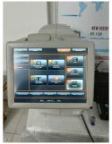
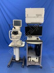
Topcon 3D OCT-1000 Mark 2 OCT system S/N: 203051 Year of Manufacture: 2008 Software version OCT: 2.02 Workstation including monitor, keyboard, mouse Software version workstation: 3.51.003.03 ... view more
Asking Price:
€2,850 EUR
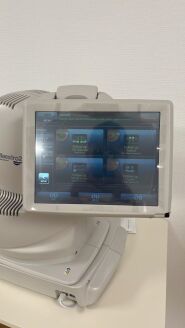
3D OCT Image Topcon’s proprietary FastMap software pioneered the 3D visualization of OCT data, providing another dimension of clinical information, thereby enhancing the understanding and... view more
Asking Price:
€9,500 EUR
Factory Refurbished Windows 10 all in one computer Includes Imagenet 6 Six months warranty No table... view more
- topcon oct-1 maestro 1 in very good conditions. - Which accessories are included! -PLEASE LET ME KNOW IF YOU NEED MORE... view more

very good condition... view more
Asking Price:
$15,000 USD

very good condition... view more

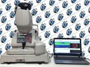
TOPCON 3D OPTICAL COHERENCE TOMOGRAPHY Year of manufacture 2015 Components - 3D OCT-1 Maestro (Application version 1.4.0.47671) - HP Z230 console - I-O DATA LCD-MF223EWR-B4 monitor - EPSON... view more
Topcon 3D OCT-1 Maestro with Anterior Segment attachment (HA-2) Input voltage: 100v~ ... view more
OCT (Optical Coherence Tomography) is a medical imaging technique that uses light waves to take cross-sectional images of the eye. OCT equipment is used to diagnose and monitor a variety of eye conditions, such as age-related macular degeneration, glaucoma, and diabetic retinopathy. It is also used to assess the health of the retina and the choroid.
The price range for OCT equipment varies depending on the type of machine and the features it offers. For example, a basic OCT machine may cost around $20,000, while a more advanced machine may cost upwards of $100,000. Some of the top manufacturers of OCT equipment include Zeiss, Topcon, and Heidelberg. These companies offer a wide variety of OCT machines, ranging from basic models to more advanced models that offer a variety of features.


 61
61