by
Brendon Nafziger, DOTmed News Associate Editor | September 23, 2009

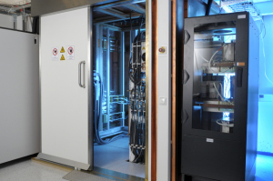
The MPI prototype,
shown here,
promises to be a faster,
stronger imaging modality
Philips has made an arrangement with German firm Bruker to manufacture its magnetic particle imaging (MPI) scanners -- a new imaging modality that promises greater resolution and speed, according to a statement released today by the companies.
The announcement of the memorandum of understanding -- a non-legally binding statement -- to make the scanners came at the World Molecular Imaging Congress in Montreal.
"We've signed the MOU. The next step would be to sign the agreement which would happen before the end of the year," Steve Klink, a spokesman for Philips, tells DOTmed News, "at which point we will determine the R&D roadmap."


Ad Statistics
Times Displayed: 3029
Times Visited: 33 Fast-moving cardiac structures have a big impact on imaging. Fujifilm’s SCENARIA View premium performance CT brings solutions to address motion in Coronary CTA while delivering unique dose saving and workflow increasing benefits.
MPI is a modality Philips began developing in 2001, which measures the magnetism of iron-oxide nanoparticles. These nanoparticles are the contrast agents injected in a subject. When exposed to a changing, or oscillating, magnetic field, the iron-oxide nanoparticles become magnetized and send out a magnetic signal. The scanner can then detect this signal, but can't pinpoint the exact location of the particles. To do that, it covers most of the particles except a tiny spot in the center with another magnetic beam which forces them to move in a fixed direction, making them invisible to the scanner. But the particles in the center can freely oscillate, and get picked up by the scanner. By moving this oscillating zone over every point in the viewing area, it can create a three-dimensional image.
Last March, Philips published a study in the journal Physics in Medicine and Biology demonstrating a prototype MPI with a 3 cm bore.
"The strong point is speed," Klink says of the modality. In the study, the MPI was able to image the heart of a mouse -- 5 mm in size, and beating at a blistering 240 beats per minute. "We were able to accurately capture the cardiovascular activity of that mouse," Klink says.
Currently, Philips hopes to go into the pre-clinical market, and has already developed, with German grant money, another prototype with a 12.5 cm bore that can be used on rabbits in cardiovascular studies.
Eventually, Klink says, Philips hopes to create a 40 cm or 60 cm bore machine for humans, but first the deal for pre-clinical equipment has to be sealed with Bruker.

