by
Lauren Dubinsky, Senior Reporter | June 15, 2015

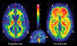
2-Amyloid imaging
with GE’s Vizamyl
From the June 2015 issue of HealthCare Business News magazine
The U.S. molecular imaging market is expected to remain steady at around $1.5 billion through 2020, but the global market is projected to reach $2.2 billion, growing at a compound annual growth rate of 3.3 percent, according to a new RnRMarketResearch report. Part of the reason U.S. PET/CT sales have been stagnant for the past couple of years have to do with hospitals’ quest to cut costs. “Customers are more conscientious of pricing,” says Robert Brait, PET national product manager for Siemens Healthcare. “When evaluating to spend a couple of million dollars on a piece of equipment, health care organizations will also have to evaluate patient volumes and how they can best treat their patient community.”
As a result, many communities, especially the smaller ones, are sharing one PET/CT scanner in a cancer center. There has also been a resurgence in mobile PET/CT, mostly in rural communities, in an effort to service a large demographic area with one mobile system. But despite flat sales, PET/CT, as well as SPECT, SPECT/CT and PET/MRI have undergone a lot of technological advancement in recent years. Quantification is now the standard for PET/CT imaging and it’s starting to gain interest in SPECT/CT imaging. Meanwhile PET/MRI is beginning to be used for clinical applications.
The changing face of PET/CT


Ad Statistics
Times Displayed: 2869
Times Visited: 27 Fast-moving cardiac structures have a big impact on imaging. Fujifilm’s SCENARIA View premium performance CT brings solutions to address motion in Coronary CTA while delivering unique dose saving and workflow increasing benefits.
In the past, CT was the only modality used for radiation therapy planning (RTP) but there is now a growing interest in using PET/CT instead. “By adding PET to the CT scan you combine the anatomic detail of a premium CT with precise metabolic information of PET to detect, characterize and monitor even the smallest cancer lesion with reproducible quantification to support more cost-effective treatment,” says Brait.
He adds that CT will only show cancer activity once the tissue has been damaged, which could be two years after it had started to form, whereas PET shows activity at the cellular level before tissue damage even occurs. Dr. Mark Winkler of Steinberg Diagnostic Magnetic Imaging in Nevada is using his recently purchased Toshiba America Medical Systems Celesteion PET/CT for RTP. “For the first time we have a way to do treatment planning not only with the anatomic landmarks of CT but with the physiological information provided by the PET,” he says.
The system, which received FDA approval in November, utilizes an 88 centimeter PET bore that makes it ideal for the range of positions that RTP requires. Patients can be scanned with their arms over their head or with their breast down, hanging freely, if they have breast cancer.

