From the November 2017 issue of HealthCare Business News magazine
Hopefully, by removing references that a particular room is defined as being a “minimum of xxx square feet,” planners, design professionals and radiology providers will engage in discussions around:
- What piece of equipment will the room need to accommodate?
- What additional equipment, material, components or supplies ought to be in the room?
- What clinical procedures may be performed in the room, and what are the space needs for those procedures?
- How might all of these (equipment, support and clinical applications) shift over time, and what are those space needs implications?


Ad Statistics
Times Displayed: 109945
Times Visited: 6642 MIT labs, experts in Multi-Vendor component level repair of: MRI Coils, RF amplifiers, Gradient Amplifiers Contrast Media Injectors. System repairs, sub-assembly repairs, component level repairs, refurbish/calibrate. info@mitlabsusa.com/+1 (305) 470-8013
While the FGI design standard can not direct communication between planners and clinical staff, one of the intentions behind stripping out explicit square footage requirements is to prompt exactly this type of dialogue. Fostering this type of dialogue with respect to room sizes becomes even more important when we look at the much larger structural change to the way that the standard looks at imaging service spaces.
Taking intended utilization into design consideration
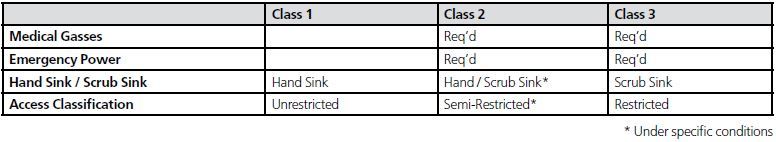
In prior editions of the FGI guidelines, a CT was a CT, irrespective of its use. There was little in the code to distinguish between a CT scanner in a physician practice’s office, one in an outpatient imaging center or the CT located in an emergency department. Despite significant differences in the level of acuity or intervention required among these different settings, design criteria were previously focused on the hardware much more so than the use of it. This is changing. The 2018 edition of the FGI guidelines introduces an intervention/acuity classification system, and class – as opposed to hardware – is the principal driver of design criteria for imaging spaces.
The new structure introduces three classes, applicable to all medical imaging devices. These new classes roughly correspond to popular understandings of “diagnostic imaging” (Class 1), “interventional imaging” (Class 2) and “intraoperative imaging” (Class 3). Class 1 should be understood to be patients not requiring significant clinical support or monitoring. Class 2 represents image-guided minimally invasive therapies or diagnostics, including angiography and MR-guided breast biopsy. Class 3 is the realm of full surgical interventions that are aided with imaging equipment.
Under this classification scheme, our prior hypothetical CT scanner would have substantially different design criteria depending on the way in which it was planned to be used. The chart on this page shows a simplified version of how some of the design requirements would change for a CT scanner suite, depending on the planned usage of the device.