by
Lauren Dubinsky, Senior Reporter | February 15, 2017
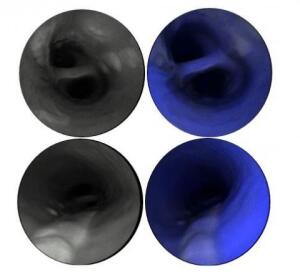
Images demonstrate multiple
atherosclerotic lesions with very
low fluorescence in the blue
spectrum in comparison to the
surrounding healthy artery
Courtesy of Michigan Medicine
In order to determine a patient’s risk of suffering a stroke or heart attack, physicians need a clear view of the carotid artery. Researchers at Michigan Medicine leveraged an existing laser-based medical camera to do what conventional radiological techniques couldn’t.
“I decided to look directly at the inner surface of arteries to identify at-risk plaques and ruptured lesions missed by conventional diagnostic modalities,” Dr. Luis Savastano, resident neurosurgeon at Michigan Medicine, told HCB News.
For every 10 patients that suffer a stroke, physicians are unable to pinpoint the cause in three or four of them, according to Savastano. It’s often suspected that ruptured plaque in the carotid artery is to blame, but it’s difficult to prove.


Ad Statistics
Times Displayed: 346433
Times Visited: 21062 MIT labs, experts in Multi-Vendor component level repair of: MRI Coils, RF amplifiers, Gradient Amplifiers Contrast Media Injectors. System repairs, sub-assembly repairs, component level repairs, refurbish/calibrate. info@mitlabsusa.com/+1 (305) 470-8013
The scanning fiber endoscope was originally developed by a mechanical engineering research professor at the University of Washington named Eric Seibel. It was designed to detect cancer in the early stages by imaging cancer cells that cannot be seen with clinical endoscopes.
Savastano and his team decided to use SFE to predict patients who are at risk for cardiovascular events, because it generates very high-resolution images and is flexible enough to navigate safely complex geometries.
SFE illuminates tissue with multiple laser beams, and digitally reconstructs high-definition images to determine the severity of the plaque build-up in the arteries. Physicians can identify and monitor the biological markers that make a plaque unstable and at risk for rupture.
They can then spot the individuals who would benefit the most from preventive treatment before symptoms appear. The plaque-specific data can be used to help physicians adjust the intensity of treatment, and the endoscope can guide stent placement and release drugs and biomaterials during therapeutic interventions.
Savastano said that PET and MR imaging are likely more convenient as initial screening in the high-risk population, but higher-resolution diagnostic approaches are often needed to detect the key markers of plaque vulnerability with high specificity and spatial resolution.
The proof-of-concept results were published in the journal
Nature Biomedical Engineering. The research is still in the preclinical stage.
Savastano and his team are continuing to conduct research to better understand the value and limitations of this technology. More research is needed about the association between high-risk plaque characteristics and clinical events.
“It is important to mention that despite major advancements in imaging modalities, we have yet to identify which biological and structural features of plaques are useful for guiding management and preventing cardiovascular events,” he added.


