by
Lauren Dubinsky, Senior Reporter | September 13, 2017
The 3-D anatomy of cardiac conduction
system in the intact human heart
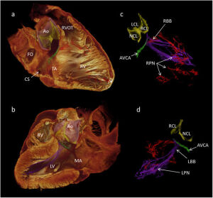
A new contrast-enhanced micro-CT imaging technique developed by Aarhus University may one day provide heart surgeons with 3-D reproductions of the cardiac conduction system.
An article was published in the journal
Scientific Reports in collaboration with researchers at Liverpool John Moores University, University of Manchester and Newcastle University that summarized the clinical and research implications.
"[This] uses similar technology to a clinical CT scanner, but allows for much higher spatial resolution," Robert Stephenson of Aarhus University, told HCB News. "Using an iodine-based contrast agent, which is taken up deferentially by different tissue types, we can identify the specialised tissue of the conduction system from the surrounding connective tissue and myocardium."


Ad Statistics
Times Displayed: 346433
Times Visited: 21062 MIT labs, experts in Multi-Vendor component level repair of: MRI Coils, RF amplifiers, Gradient Amplifiers Contrast Media Injectors. System repairs, sub-assembly repairs, component level repairs, refurbish/calibrate. info@mitlabsusa.com/+1 (305) 470-8013
This technique is useful for procedures that involve positioning a heart valve prosthesis a few millimeters from the conduction system. It also has implications for the treatment of atrial fibrillation, in which knowledge of the conduction system becomes very important.
Treating atrial fibrillation requires the administration of medicine or electrophysiological stimulations, and in rare instances a pacemaker needs to be implanted to restore normal pump function.
"The technique has potential for characterising the 3-D disposition of the conduction system in different disease states and malformations," said Stephenson. "It is this information which will be used to inform surgeries."
The researchers believe that this technique could also be used to train medical students. The 3-D visualization of the cardiac conduction system can help them learn about its complex relationship with the heart anatomy and function.
To date, the research team has only presented 3-D visualization of a healthy human heart, but they plan to focus on diseased hearts next. That would include patients suffering from congenital heart disease and the elderly population.


