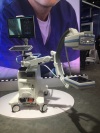
The system can recognize the device specs being considered for use, such as balloons or stents, then virtually overlay the device on the virtual angiographic image of the vessel for proper sizing in both length and diameter. The demo allowed the user to bring up a holographic image of a 3-D model of the vasculature, which one could then rotate to see the X, Y, and Z axis perspectives without ever moving away from the table or breaking the sterile field.
I believe that the ability to consider all the imaging, monitoring, device data, and 3-D modeling, as well as live procedure imaging – not just from memory, but seen in the virtually augmented presentation – offers physicians and patients the information and images to more efficiently and accurately make decisions in “real time”.


Ad Statistics
Times Displayed: 112999
Times Visited: 6736 MIT labs, experts in Multi-Vendor component level repair of: MRI Coils, RF amplifiers, Gradient Amplifiers Contrast Media Injectors. System repairs, sub-assembly repairs, component level repairs, refurbish/calibrate. info@mitlabsusa.com/+1 (305) 470-8013
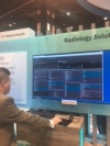
Philips most decidedly is not the only vendor aggressively pursuing the integration of Artificial Intelligence (AI), Machine Learning (ML), and/or Deep Learning (DL). These terms are somewhat synonymous, or certainly overlap, in the realm of virtual reality. They were a major focus of many vendors and presentations during RSNA. It is important to note that I use Philips only as a working demonstration as to what is possible, and not as an endorsement of its technology over that of any other vendors. Philips simply had a functional display that demonstrated the tremendous potential of AI/ML/DL in a manner that, I believe, supports Dr. Zerhouni’s first principle: “What will be is already here.”
The use of augmented reality is taking what is already here and possible with MRI, CT, IVUS, biological testing, 3-D reconstruction and modeling, ultrasound, and ultrasound-based guidance, and presenting the information, data, and imaging in “real time” for the physician to remain focused on the patient procedure in progress. With this information the physician can make decisions based on a wide array of other procedures and imaging to optimize decisions and patient care.
I believe my initial question, “A New View or Just a Different View?” is answered, as it is both!
About the author: Tom Watson joined MD Buyline in 1986 and has over 40 years of experience in the field of cardiovascular medicine. He started his career as a staff technologist at West Jefferson Medical Center in Marrero, La. He spent two of those years as a staff technologist and the remainder as administrative and technical director of cardiology. His clinical experience and training include all diagnostic noninvasive cardiac modalities, including echocardiography, stress-testing Holter and ECG. His invasive experience encompasses diagnostic cardiac catheterization, PTCA and electrophysiology. He also has experience with critical care and intensive care monitoring, as well as cardiac rehabilitation applications. As a senior clinical analyst at MD Buyline, Watson is the primary analyst for cardiology, which includes interventional angiography (cardiac, vascular and neurological), electrophysiology imaging and monitoring and cardiology PACS and information systems. He provides secondary support for noninvasive cardiology. He also provides cross-coverage to related areas of radiology.
Back to HCB News