From the November 2020 issue of HealthCare Business News magazine
The scanner is expected to benefit community, rural and private hospitals, as well as urgent care centers, and orthopedic and large radiology practices, all of which would use different components of the system from one another based on their diverse needs.
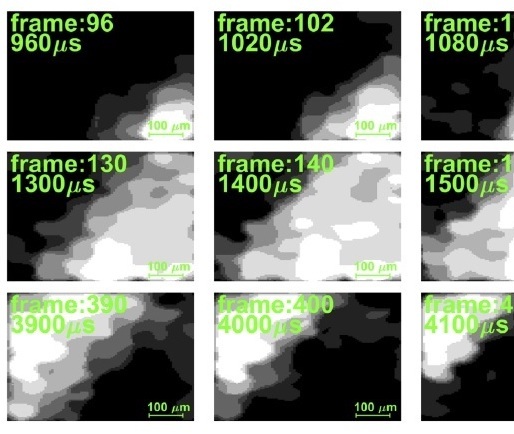
Researchers use X-ray to capture fast moving organs in hi-res
A novel technique announced by researchers in August could one day enable providers to
capture the motion of fast moving biological processes previously unobservable with X-ray imaging.


Ad Statistics
Times Displayed: 72233
Times Visited: 2363 Ampronix, a Top Master Distributor for Sony Medical, provides Sales, Service & Exchanges for Sony Surgical Displays, Printers, & More. Rely on Us for Expert Support Tailored to Your Needs. Email info@ampronix.com or Call 949-273-8000 for Premier Pricing.
The inspiration behind the technique is a non-traditional method known as ghost imaging, which not only improves imaging speeds but reduces radiation exposure to patients.
“The high frame rate that the ghost imaging technique has demonstrated in the present work should be beneficial for medical procedures that require information on moving organs, the most prominent one being the heart,” lead researcher, professor Sharon Shwartz, and Ph.D. student Mr. Or Sefi told HCB News. “Imagine that you can get a high-resolution high-frame movie of the moving heart. This way you get not only the information on its anatomy but also on its function.”
The researchers used the approach to create a movie of a blade rotating at 100,000 frames per second. They first used standard sandpaper mounted on motorized stages to create the reference beam and form a random pattern that was recorded with a high-resolution, slow frame-rate pixelated X-ray camera.
Shwartz and Sefi say the technique can be used with any X-ray source and can enable clinicians to use X-rays to measure fast dynamics outside the lab. They are continuing to make improvements to the overall system and the image reconstruction algorithm for greater resolution and shorter measurement times.
FDA gives nod to KA Imaging's dual-energy detector
In September the FDA gave KA Imaging clearance to distribute the Reveal dual-energy, single exposure X-ray detector, which can
differentiate between bone and soft-tissue without motion artifacts in one shot.
Reveal can provide an unobstructed view of the lungs, which can help in visualizing pneumonia, fractures, catheters, and masses with high sensitivity. Its use of very high detective quantum efficiency simultaneously allows for low radiation exposure and high-quality soft tissue and bone images.