Sponsored Content | December 21, 2023

The first dedicated breast coil was introduced more than 30 years ago. Since that time, breast MR has proven its value as a breast imaging modality. MRI can detect lesions not seen on mammography and ultrasound, and can effectively evaluate the breast when density, implants or post-surgical scarring interfere with conventional mammography. Breast MRI is also used to evaluate the extent of disease as part of preoperative assessment in women with newly diagnosed breast cancer, because it enables delineation of the primary tumor, assessment of the lymph nodes, diagnosing multicentric and contralateral disease, and evaluating local nodal metastases and chest wall involvement. During treatment, MRI can be used to assess a patient's response to neoadjuvant chemotherapy.
While breast MRI will likely never replace mammography - at least in our lifetime - as first line imaging for most patients, its use as a screening tool has also grown.
One trend is toward screening tailored to an individual woman's risk of developing breast cancer. This more personalized approach may include mammography, ultrasound and MRI as well as ultrasound tomography or abbreviated breast MRI.
This has already been implemented for those at greatest risk. The American Cancer Society and American College of Radiology recommend that women at substantially increased risk of breast cancer -- defined as a 20% or greater lifetime risk of developing breast cancer --be screened annually with contrast-enhanced breast MRI in addition to annual mammography starting at 25 years, as MRI remains the most sensitive imaging modality available for the detection of breast cancer. This group includes women with the BRCA gene, those exposed to chest radiation at a young age, women diagnosed with breast cancer prior to age 50 and also those with dense breasts, who have a 4.6-fold increased risk of developing breast cancer when compared to average risk women. MRI is valuable for this population because in women with dense breasts, the sensitivity of mammography is decreased.
Factors limiting growth
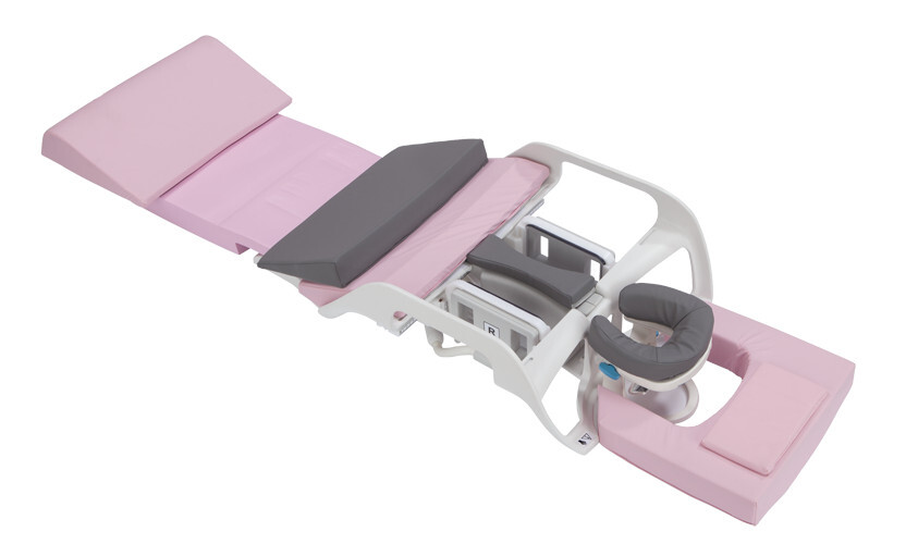
However, several factors have limited more widespread use of breast MRI. As with other MR exams, these factors are: cost, the availability of MR systems, and exam length.
Exam length
This last area - exam length - is where we have control to create a screening or abbreviated protocol using the mask and first post-contrast 3D. In addition, increased channel count of coils results in higher SNR, use of higher acceleration factors enables higher resolution in the same time, and 3D sequences with fat suppression are now possible. Even if you believe a T2 Fast spin echo with Fat Saturation or a STIR is needed, it is now possible to conduct a MR breast assessment in under 10 minutes.
It may be possible to reduce scan time even further! A recent study showed an abbreviated protocol of just 3 minutes was sufficient to establish the absence of breast cancer. 1
Improving workflow
Of course, when it comes to total exam length, shortening the scan time is only half of the story. To truly shorten the length of a breast MR exam, we need to improve workflow, from the moment the patient checks in until the exam is finished.
This starts with explaining the exam and reducing anxiety before the patient enters the exam room. Having the patient changed into a hospital gown and with an IV started while in the waiting room are two ways to maximize efficiency. While effective, this will require a change in staffing so that one technologist can be talking to and preparing a patient while another is running an exam, and perhaps a third is queueing up a third patient, or post-processing images.
Another way to make breast MRI more efficient is to block schedule - performing all breast exams in a designated time slot. This allows techs to get into a rhythm, with no need to change coils between patients and use of the same protocols for all patients in a certain time block.
Shorter exams may also allow hospitals to serve more patients, which could lead to increased patient satisfaction and revenue.
The future
So, what can we expect in the future? Look for the combination of abbreviated protocols and acceleration techniques to significantly shorten exam length, shorter dynamics using different k-space filling techniques to acquire more kinetic information in the first 90 seconds, and different acceleration methods. Watch for hospitals to get creative with staffing and scheduling to enhance efficiency.
We also anticipate that new coils with even higher channel counts will increase SNR and allow even greater imaging quality or shorter scan times. And, as with so many things these days, increased use of artificial intelligence will bring even more efficiency to image acquisition, reconstruction, post-processing and diagnosis.
________________________________
1 Kuhl C, Schrading S, Strobel K, Schild H, Hilgers R, Bieling H. "Abbreviated Breast Magnetic Resonance Imaging (MRI): First Postcontrast Subtracted Images and Maximum-Intensity Projection - A Novel Approach to Breast Cancer Screening with MRI." Journal of Clinical Oncology. August 1 2014;32(22):2304-14.
About Dunlee
Dunlee has over 100 years' experience in developing, producing and integrating innovative components for imaging systems. Serving the OEM market, Dunlee offers a comprehensive portfolio of reliable X-ray tubes, high voltage generators, detectors and product packages for CT, as well as solutions for interventional radiology and MRI and additive manufacturing solutions with tungsten. It offers support during development and throughout the product lifecycle, contributing to its customers' efficient production and go-to-market strategies. Visit www.dunlee.com to learn more.
Back to HCB News


