by
Olga Deshchenko, DOTmed News Reporter | May 16, 2011
From the May 2011 issue of HealthCare Business News magazine
To better serve women when mammography isn’t enough, several companies and researchers are bringing ultrasound technology into the breast imaging realm.
And while some market their devices as a means to ensure a mammogram didn’t miss anything, others hope their innovations challenge, if not entirely replace, current breast screening technology.
Now on the market


Ad Statistics
Times Displayed: 136729
Times Visited: 7931 MIT labs, experts in Multi-Vendor component level repair of: MRI Coils, RF amplifiers, Gradient Amplifiers Contrast Media Injectors. System repairs, sub-assembly repairs, component level repairs, refurbish/calibrate. info@mitlabsusa.com/+1 (305) 470-8013
In early April, at a breast density education seminar at its headquarters, U-Systems announced its partnership with Are You Dense.
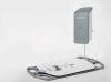
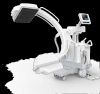
U-Systems manufactures the somo•v Automated 3-D Breast Ultrasound system, which has Food and Drug Administration approval for diagnostic use. If a physician detects an abnormality, the device can be used to characterize it, says Ronald Ho, the company’s CEO and president.
The technology behind the system was developed by U-Systems founder Bob Wong more than a decade ago. Wong understood that mammography was not very sensitive with dense breast tissue and that ultrasound did better in such cases. “But he also realized that a handheld scanning of the breast was not viable for a screening method,” says Ho.
A screening with a handheld ultrasound is lengthy (about 30 to 40 minutes) and can’t be replicated. To counter these disadvantages, Wong came up with a method that uses a large, 15-centimeter transducer and allows for an automated process – just as with mammography, the acquisition is mechanical.
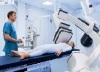
To obtain the images, a technologist positions the ultrasound scanner on the patient’s breast while the woman is on the exam table. The acquisition process takes less than 15 minutes. The device also enables radiologists to review the images in a batch mode – the software ensures quick access to the results, says Ho.
“It’s really taking the very effective imaging modality of ultrasound and applying it in a reproducible way so it can be a real screening process,” he says.
One of the biggest challenges U-Systems is facing is overcoming the lack of knowledge around breast density. Ho says the issue is a “double whammy,” as the most popular breast screening technology doesn’t handle dense tissue well and women with dense breast are at a higher risk for cancer.
Today, about 55 of U-Systems’ devices are in use worldwide. The company is also seeking FDA approval for its system for adjunctive use in breast cancer screening.
U-Systems is currently sponsoring the prospective Somo•Insight Clinical Study, designed to see if digital mammography coupled with its ultrasound device is more sensitive than a routine screening mammogram alone in finding cancers in women with dense breast tissue. So far, more than 13,000 women have participated in the study nationwide.