by
Olga Deshchenko, DOTmed News Reporter | May 16, 2011
From the May 2011 issue of HealthCare Business News magazine
“Our performance specification is to see 100 percent of lesions greater than 5 millimeters,” says Robinson. “We have seen lesions as small as 2 millimeters.”
In the university
Many promising inventions that use ultrasound technology to overcome the shortcomings of mammography are also brewing in educational facilities across the country.


Ad Statistics
Times Displayed: 78539
Times Visited: 2794 Ampronix, a Top Master Distributor for Sony Medical, provides Sales, Service & Exchanges for Sony Surgical Displays, Printers, & More. Rely on Us for Expert Support Tailored to Your Needs. Email info@ampronix.com or Call 949-273-8000 for Premier Pricing.
Thomas R. Nelson, a professor of radiology and bioengineering at the University of California, San Diego, is working on what he calls a volume breast ultrasound scanner.
In an e-mail to DOTmed News, Nelson, who has been doing breast cancer detection research for the past 15 years, said he anticipates that his device will ultimately do just as good, if not better than mammography in detecting lesions.
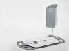
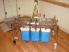
The VBUS acquires images while a woman’s breast is in a heated water bath. The data is then reconstructed to produce high-resolution tomographic images. “We also measure other acoustic parameters to better understand the breast tissue properties that ultimately will better characterize cancer,” said Nelson.
The most difficult part has been the design of the specialized hardware, necessary to obtain high quality data, and the invention of new analysis strategies, he said. The VBUS is currently in preliminary clinical trials.
And a professor of biomedical engineering at the University of Southern California in Los Angeles is working on the development of a device he hopes can push mammography off its pedestal as the leading modality for early detection and breast cancer screening.
In March, Vasilis Marmarelis presented on the potential of his Multimodal Ultrasound Tomography technology at the European Congress of Radiology in Vienna.
In a clinical study of 32 volunteers, the MUT device correctly identified 27 malignant and 19 benign lesions, all of which were confirmed by biopsies.
Since then, the study ha been expanded to 64 volunteers, presenting 42 malignant and 51 benign lesions which ranged in size from 1 to 38 millimeters. All lesions were detected and correctly differentiated by MUT.
Marmarelis’ technology can offer a reliable way to detect breast cancer early, when lesions are roughly 5 millimeters, and even as small as 1 millimeter in size, a dimension that “cannot be achieved with high specificity by any other existing technology,” he says.
Marmarelis also says his technology is capable of characterizing lesions noninvasively, eliminating the need of biopsies. (In his study, correct noninvasive differentiation was achieved in all cases to date.)