by
Carol Ko, Staff Writer | May 03, 2013
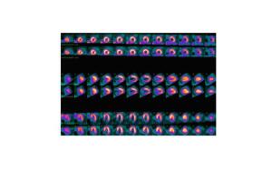
A new PET tracer
for myocardial perfusion
is one step closer
to market release.
A new PET tracer may now be one step closer to shaking up the nuclear imaging market. Lantheus Medical Imaging announced this week that the first of two Phase 3 studies for flurpiridaz F 18, a tracer used in PET imaging, has met the criteria for completion.
The tracer is designed to assess myocardial perfusion, or blood flow to the heart, using PET (Positron Emission Tomography) scans.
The test helps diagnose patients with known or suspected coronary artery disease, the leading cause of death in men and women in the United States. It affects 16.8 million Americans, each year killing over half a million.


Ad Statistics
Times Displayed: 3341
Times Visited: 6 Stay up to date with the latest training to fix, troubleshoot, and maintain your critical care devices. GE HealthCare offers multiple training formats to empower teams and expand knowledge, saving you time and money.
Currently, most myocardial perfusion tests are done with SPECT, not PET, but that may change with the entrance of new PET tracers such as flurpiridaz F 18.
Supply chain woes around SPECT may also boost this radiopharmaceutical's adoption, if approved. The most common radiopharmaceutical used for SPECT myocardial perfusion, technetium-99, requires radioisotopes that are made in only five aging nuclear reactors around the world.
Phase 2 trials showed that the PET agent may enable better image quality than SPECT perfusion imaging and reduce attenuation, or loss of image accuracy. To date, approximately 900 subjects have been imaged with flurpiridaz F 18.
"When we start our second Phase 3 trial will depend on the timing of successfully completing our ongoing strategic partner discussions," Simon Robinson, vice president of research and pharmaceutical development at Lantheus, told DOTmed News.


