by
Carol Ko, Staff Writer | August 13, 2013
From the August 2013 issue of HealthCare Business News magazine
But the gamma knife had some worrisome drawbacks for Lockwood, including swelling of the brain and lower rates of efficacy. When Lockwood found out about new MRI-guided laser ablation, it seemed like the noninvasive option with the fewest drawbacks.
The treatment was pioneered by surgeons at the Texas Children’s Hospital, where the first surgeries were performed by Dr. Daniel Curry, director of pediatric surgical epilepsy and functional neurosurgery and Dr. Wilfong. Both doctors had noted that MRIguided laser technology was already being used to treat certain tumors.


Ad Statistics
Times Displayed: 45175
Times Visited: 1385 Keep biomedical devices ready to go, so care teams can be ready to care for patients. GE HealthCare’s ReadySee™ helps overcome frustrations due to lack of network and device visibility, manual troubleshooting, and downtime.
“We thought, ‘why couldn’t we use this technology approved for tumors to treat epilepsy?’” said Wilfong. He and Curry met with the manufacturer of this technology, Visualase, and evaluated a series of patients who could potentially benefit from this procedure. Upon gaining approval through an institutional review board, they performed their first case three and a half years ago.
Since then, the surgeons have performed almost 30 cases, and more than 150 cases have been performed around the nation. “The vast majority of patients, instead of spending five to seven days in the hospital after a craniotomy, go home the next day,” says Wilfong.
Pins and needles
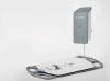
The procedure does pose certain technical challenges for doctors performing the surgery: the most critical component of the procedure’s success is the precise placement of the laser probe —to the millimeter, experts say.
Doctors start by performing an MRI scan on the patient that shows the vessels on the brain. Then, they place a frame on the skull with a coordinate system. A second scan is combined with the first to show exactly where and at what angle to place the probe. An incision the size of a pinhole is made in the head and the probe is bolted to the skull.
The patient is then sent to the MRI suite, where the probe is heated up and the laser zaps abnormal brain tissue. After the treatment is completed, doctors remove the probe and close up the incision. Patients are normally ready to leave the hospital the next day.
Border Control
No matter what technique neurosurgeons use to target abnormal brain tissue, there’s always a penumbra of tissue around the treated area called a drop-off — where tissue has been damaged but not killed — creating areas of the brain that are not totally dead, but not fully functioning either. In that regard, lasers provide an advantage for surgeons because their drop-off is very sharp. The greater the drop-off is, the greater the benefit to the patient.