by
Lauren Dubinsky, Senior Reporter | August 11, 2014
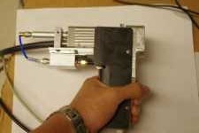
Researchers from Washington University in St. Louis created a new hand-held device that might change the way physicians diagnose and treat melanoma. The device was described in a paper published yesterday in The Optical Society's journal, Optics Letters.
It's called a photoacoustic microscope and works by measuring how deep a melanoma tumor is. A laser beam shines into the skin on the tumor and melanin absorbs the light and the energy is transformed into high-frequency acoustic waves.
The acoustic waves don't scatter as much as light when it travels through the skin so they're able to penetrate deeper. Since the tumor cells produce more melanin than the healthy skin cells around it, the acoustic waves can map the whole tumor with high resolution.


Ad Statistics
Times Displayed: 1227
Times Visited: 6 Fast-moving cardiac structures have a big impact on imaging. Fujifilm’s SCENARIA View premium performance CT brings solutions to address motion in Coronary CTA while delivering unique dose saving and workflow increasing benefits.
There's a detector on the device that can then turn the acoustic signal into a 3-D image on a screen.
The traditional ways — high-resolution optical techniques — to diagnose and determine treatment don't measure the tumor that well because they don't transform the light into acoustic waves. Other method such as high-frequency ultrasound, MR and PET use acoustic waves but ultrasound doesn't have enough image contrast and MR and PET have inadequate resolution.
In order to determine if the tumor is cancerous, a biopsy has to be performed to remove part of the tumor. If the tumor depth is not accurately measured before the biopsy and the surgeon finds out during the procedure that the tumor is deeper than they thought, the patient may have to undergo another surgery.
"The surgical treatment plan is dependent upon determining the depth of tumor invasion into the skin, and this cannot be adequately determined when only part of the lesion has been sampled," Lynn Cornelius, one of the authors of the study, wrote to DOTmed News. "This device will potentially allow us to scan the remaining tumor on the skin to determine the depth of invasion and plan surgical management appropriately."
The previous desktop version of the device shone the laser directly onto the tumor but so much light was being absorbed and not much of it penetrated the tumor's lower layers. The newer hand-held device distributes the light around and below the tumor, which produces a bright image of the tumor's bottom and gives an accurate measurement of its depth.
The researchers have tested it on artificial tumors made of black gelatin and also on live mice. They proved that it can accurately measure the depths of tumors but now it needs to be proven in human patients.
They are currently conducting a study with 25 human patients at the university and if it proves effective then it will become widely available.
The tool will mostly be used for improving the planning and preparation for surgeries but since it measures the tumor's whole volume, it can help in other ways. If the researchers can find out how the volume of the tumor is related to cancer outcomes then the device could provide physicians with a new type of measurement for prognosis.

