by
Barbara Kram, Editor | October 24, 2006

Miniature endoscope uses micro optics
BOSTON - Massachusetts General Hospital (MGH) researchers have developed a new type of miniature endoscope that produces three-dimensional, high-definition images, which may greatly expand the application of minimally invasive diagnostic and therapeutic procedures. In the October 19 issue of Nature, the team from the Wellman Center for Photomedicine at MGH describes their prototype device and a demonstration of its use in a mouse model.
"This new ultraminiature endoscope is the first to allow three-dimensional imaging of areas inside the body, " says Guillermo Tearney, MD, PhD, of the MGH Wellman Center, the report's senior author. "Its ability to go places that other imaging tools cannot reach opens new possibilities for medical diagnosis and eventually treatment."
Standard miniature endoscopic devices - which give physicians access to hard-to-reach internal organs and structures - utilize bundles of optical fibers to supply light to and transmit images from the areas of interest. Larger endoscopes that use image sensors to produce high-quality, two-dimensional images can be a centimeter or more in diameter. Existing miniature endoscopes using smaller fiber bundles may be more flexible but have difficulty producing high-quality images.


Ad Statistics
Times Displayed: 45539
Times Visited: 1299 Ampronix, a Top Master Distributor for Sony Medical, provides Sales, Service & Exchanges for Sony Surgical Displays, Printers, & More. Rely on Us for Expert Support Tailored to Your Needs. Email info@ampronix.com or Call 949-273-8000 for Premier Pricing.
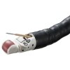
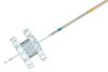
The new device developed at MGH-Wellman uses a technology called spectrally encoded endoscopy (SEE). Multicolored light from a single optical fiber - introduced through a probe about the size of a human hair - is broken into its component colors and projected onto tissue, with each color illuminating a different part of the tissue surface. The light reflected back is recorded, and the intensity of the various colors decoded by a spectrometer, which analyzes the wavelengths of light. Another device called an interferometer, which calculates structural information based on the interaction between two waves of light, provides the data required to create three-dimensional images.
To demonstrate the device's application in a live animal, the researchers used the system to image metastatic ovarian tumors on the abdominal wall of a mouse. The SEE probe was passed into the abdominal cavity through a fine-gauge needle. The resulting three-dimensional image showed several raised areas of tumor nodules, the presence of which was confirmed by histologic analysis of the tissue.
"The most important feature of this new endoscope is the ability to obtain three-dimensional images, something we don't believe is offered by any commercially available miniature endoscope system," says Dvir Yelin, PhD, first author of the Nature paper. "While the image resolution we achieved in this demonstration is similar to existing small-diameter endoscopes, with further optimization of the optics it is possible to obtain images with 10 times the number of pixels provided by other miniature endoscopes."