by
Gus Iversen, Editor in Chief | November 23, 2015
HCB News: What are the potential applications?
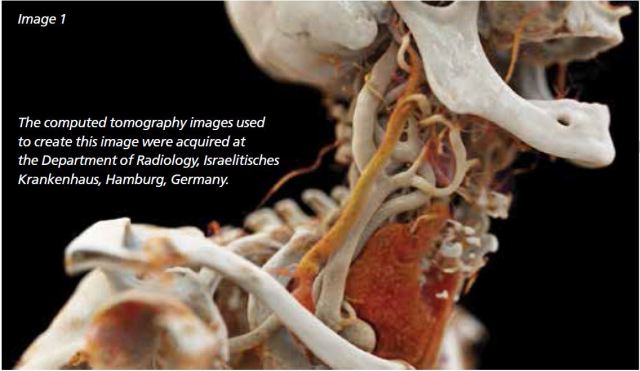
DC: This new technology has the potential to enhance clinician-patient communication. Instead of seeing traditional black and white images, if you look at Image 1 of our presentation you see a great visualization of the neck, including the carotid artery, jugular vein and thyroid gland. Suddenly, things are much simpler to understand compared to a traditional CT or MR image.


Ad Statistics
Times Displayed: 19090
Times Visited: 362 Stay up to date with the latest training to fix, troubleshoot, and maintain your critical care devices. GE HealthCare offers multiple training formats to empower teams and expand knowledge, saving you time and money
Second, it’s about revealing more human anatomy and function through medical imaging. During discussions with our key luminaries, we talked about a turning point in teaching anatomy. Today most of academic teaching is done on cadavers through dissection, but you don’t deal with the living body. The fascinating part here is thinking of the opportunity to teach about the living body via a virtual dissection facilitated by medical imaging and photorealistic visualization. Third, we focus on increasing the sensitivity of diagnostic imaging. Cinematic rendering can contribute to revealing disease in early stages.
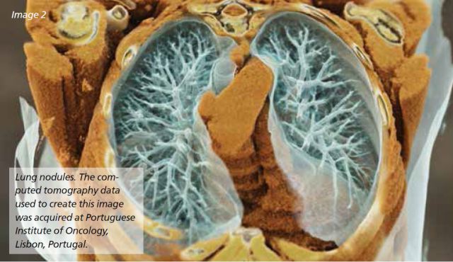
Think, for example, about enhancing the conspicuity of lung nodules in CT data. We provide an example of nodules in Image 2. Here, through the capturing of detailed image properties, you can see a much richer depth representation of data. Furthermore, we can focus on better staging, and better quantification of tumors. In general, photorealistic visualization brings value every time one needs to quantify pathologies in images. The fourth impact is about decision-making. Can we increase specificity? What is the type of disease? Can we better stratify or classify disease? Can we derive more information from the data? What is the status of implants?

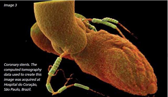
Multiple coronary stents are highlighted in Image 3. Then, No. 5, if one sees more through medical imaging, one can better guide minimally invasive procedures such as aortic stent implants, heart valve implants, and heart, liver or spine surgery. Minimally invasive surgery is developing rapidly today to minimize trauma on the patient and reduce the associated cost. Medical imaging constitutes the main source of information for guiding such procedures.
HCB News: What’s next for this technology?
DC: Personalized therapy is the next big topic, and we are addressing it on the research side. We think of obtaining information not only at the anatomical level, but also at the physiological one. Take the heart, for example. You have the heart anatomy together with information about electrical signal propagation, the dynamics of blood being pumped by the heart, and the electromechanical information on how the heart muscles function. All of this information comes together to form a comprehensive, virtual, yet personalized representation of the patient’s heart. Such models can become the base for future individualized therapies.
One last comment: all images that we present in this article are images of real, living patients. When you look at the brain, it is a living brain. The image has been created through MR, noninvasively, and yet it looks so realistic. We have much more to explore to understand the full potential of this fascinating technique of cinematic rendering. Siemens Healthcare plans to display new cinematic rendering images at its exhibit hall booth (booth #4136, South building of Mc-Cormick Place, Hall A) during RSNA.
Back to HCB News