by
Christina Hwang, Contributing Reporter | June 22, 2016

Device allows molecular
imaging system to get six
times better contrast of tumors
Credit: DOE's Jefferson Lab
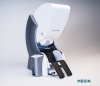
A new device called a variable angle slant hole (VASH) collimator can provide up to six times better contrast of tumors in a patient’s breast when it replaces a component in molecular breast imagers, according to developers from the Department of Energy’s Thomas Jefferson National Accelerator Facility.
Based on calculations performed by Dr. David Gilland at the University of Florida, the Jefferson team built “proof of principal” collimator prototypes, Drew Weisenberger, group leader of the Jefferson Lab Radiation Detector and Imaging Group, told HCB News.
The VASH collimator — for which two filings to the U.S. Patent and Trademark Office have been issued — is made from 49 tungsten sheets with identical square holes in each. The sheets are stacked like a “deck of cards” and have angled edges on two of its sides. The angle of the square holes can then be slanted by motors that move them by their edges, and this results in being able to change the angle of the collimator during imaging.


Ad Statistics
Times Displayed: 2436
Times Visited: 4 Stay up to date with the latest training to fix, troubleshoot, and maintain your critical care devices. GE HealthCare offers multiple training formats to empower teams and expand knowledge, saving you time and money.
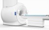
“Now you can get a whole range of angles of projections of the breast without moving the breast or moving the imager,” said Weisenberger. “You’re able to come in real close, you’re able to compress the breast, and you can get a one-to-one comparison to a 3-D mammogram.”
The new system was tested by researchers who evaluated the spatial resolution and contrast-to-noise ratio in images of a “breast phantom”, a plastic breast that had four different-sized beads that represented cancer tumors.
The beads were marked with a radiotracer and then when the collimator was used with a breast imaging system, the team found that they could get six times better contrast of the “tumors”, which could potentially reduce radiation dose to the patient while maintaining the same or better image quality.
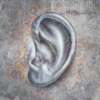
“These results really focus on the breast,” said Weisenberger. “We hope to build on this to perhaps improve the imaging of other organs.”