by
Lauren Dubinsky, Senior Reporter | July 11, 2017
Researchers at Osaka University in Japan have developed a new imaging system that can image a whole mouse brain at high spatial resolution in less than two-and-a-half hours.
The current methods for imaging a whole mouse brain at a resolution high enough to obtain useful information takes up to one week. Comparing multiple brains is necessary for understanding neurobiological function and dysfunction in brain disorders, but it’s not possible to image and analyze multiple brains with those methods.
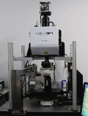
The new block-face serial microscopy tomography (FAST) technique involves spinning a disk confocal microscope with built-in a microslicer, and a method for processing image data. With this 3-D reconstruction technique, whole brains can be visualized at a resolution high enough to view individual cells and their subcellular structures.


Ad Statistics
Times Displayed: 57392
Times Visited: 1683 Ampronix, a Top Master Distributor for Sony Medical, provides Sales, Service & Exchanges for Sony Surgical Displays, Printers, & More. Rely on Us for Expert Support Tailored to Your Needs. Email info@ampronix.com or Call 949-273-8000 for Premier Pricing.
When FAST was used in conjunction with specific staining procedures, the researchers were able to visualize subcellular nuclei, vascular structures, mature oligodendrocytes, myeline sheaths, interneurons and projecting neurons throughout the whole brain.
Since FAST is such a quick imaging technique, it can also be used to image primate brains. The team was able to successfully visualize a long-range neuronal projection at a subcellular resolution in the whole brain of an adult marmoset.
The team also successfully imaged post-mortem human brains with FAST. They are hoping to identify fine morphological abnormalities in diseased human brains that were previously unknown to help in the development of effective treatments.

