by
John R. Fischer, Senior Reporter | August 29, 2017
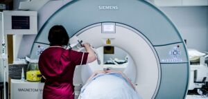
New MRI scan detects risk of
stroke in plaques (picture courtesy
of the University of Oxford)
A new type of MR scan for examining plaques might mean the difference for preventing the occurrence of stroke.
Researchers at Oxford University and surgeons at John Radcliffe Hospital have developed a noninvasive technique for analyzing and assessing if cholesterol levels in carotid arteries indicate that a person is at risk for a stroke. Their findings were published in the journal,
JACC: Cardiovascular Imaging.
“Stroke can be caused by the rupture of plaques in the neck arteries that block the blood and starve the brain of oxygen,” Luca Biasiolli, a principal investigator at the University of Oxford and one of the authors of the study, told HCB News. “Several previous studies have shown that fatty plaques with more cholesterol are more likely to break.”


Ad Statistics
Times Displayed: 346433
Times Visited: 21062 MIT labs, experts in Multi-Vendor component level repair of: MRI Coils, RF amplifiers, Gradient Amplifiers Contrast Media Injectors. System repairs, sub-assembly repairs, component level repairs, refurbish/calibrate. info@mitlabsusa.com/+1 (305) 470-8013
The standard method for assessing the risk of stroke measures the size of plaques in carotid arteries, recommending surgery for those that are deemed too big. This technique, however, may miss plaques that are not big, but are at high risk of rupturing.
The new MRI method was developed specifically to identify plaques with cholesterol levels that indicate a high risk of stroke potential, compared to ones that are stable.
To execute the project, researchers used a 3T MRI system and employed software they developed for measuring the plaques. They then performed cardiovascular magnetic resonance scans to measure the cholesterol levels of carotid plaques in 26 patients scheduled for surgery to remove their plaques.
Following the operations, the team assessed the cholesterol levels of each individual's plaque and compared these findings to the MRI scans, finding strong correlation between the two, based on individual slices and plaque average.
“Our technique can accurately measure the amount of cholesterol in plaques, as validated by analysis of samples from surgery,” said Biasiolli. “Potentially, with the necessary technical development, it could be used to measure cholesterol within plaques in other vessels, such as the aorta.”
This is not the only recent innovation in MR. A recent study found that MR could be used to predict the
suitability of epilepsy patients for surgery to stop the occurrence of seizures.
The team plans to continue studying the use of the scan on larger groups of patients. The project is supported by the British Heart Foundation Centre of Research Excellence in Oxford and the NIHR Oxford Biomedical Research Centre.
Back to HCB News


