From the October 2017 issue of HealthCare Business News magazine
This requires, however, that the therapeutic procedure is performed in the MR suite or the MR system is placed in a dedicated surgery room. The latter is an option that is explored by several institutions, but comes with very high investments and very highly skilled interdisciplinary staff.
Difficult patient access in conventional horizontal field superconductive magnets, and the need for special non-ferromagnetic therapy tools have also prevented a widespread occasional interventional use of conventional diagnostic MR systems. The interventional and therapeutic use of MR follows for the last 20-plus years the strategy to employ conventional standard high-field systems and add more or less complicated and expensive components.


Ad Statistics
Times Displayed: 109945
Times Visited: 6642 MIT labs, experts in Multi-Vendor component level repair of: MRI Coils, RF amplifiers, Gradient Amplifiers Contrast Media Injectors. System repairs, sub-assembly repairs, component level repairs, refurbish/calibrate. info@mitlabsusa.com/+1 (305) 470-8013
MR-compatible robotic systems were, for example, proposed to solve the patient access issue in combination with specially shielded in-room monitors, dedicated nitinol or plastic devices with no or little susceptibility-related imaging artifacts, and in-room optical tracking systems. These components are quite expensive and the possible therapies are still limited because of the tight space in the MR bore and the issues around surgical sterility. The robotic systems are even needed with the short-bore magnets of the newest MR generation. With a magnet length of 1.2 meters it is still 60cm (an arms length) to the center of the magnet and with a bore diameter of 70cm, with a mid-size patient placed, there is only 10-15cm of available vertical space for a therapy tool (see figure 1).
Open vertical field magnets do provide some benefits for interventional procedures, but typically have only magnetic field strength of 0.2T to 0.4T with currently only one system over 1T. Lower field strength comes with increased acquisition times, which is not good for therapy applications, and a lower image quality. On the other hand, the lower field strength also reduces susceptibility artifacts and comes with a much lower magnetic attraction force, making the surgery generally safer.
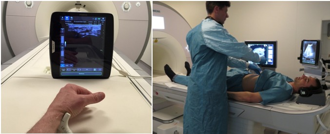
Figure 2: A tablet ultrasound of a large diagnostic imaging vendor placed only 1m away from the 3T MRI of another large imaging vendor. The ultrasound system is not approved and certified for use in the MRI suite, but direct applications under ultrasound guidance could give a new boost to performing interventional and therapeutic procedures in the MRI suite under ultrasound guidance. The picture on the right shows the cognitive fusion of MRI and US images on separate monitors.