From the October 2017 issue of HealthCare Business News magazine
So why not use the MR imaging capabilities and continue the procedure inside the imaging suite with an ultrasound system that is either cognitively fused (see figure 2) or automatically co-registered and overlaid to the just obtained MR images. Several commercially available ultrasound systems could theoretically be used even up to 1m to a 3T magnet opening (see figure 2). While these systems do not have an official MR safe label, research institutions have used these systems in phantom set-ups to rethink interventional MR procedures.
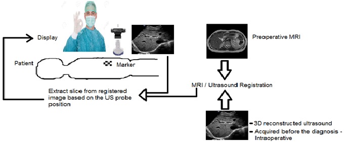
First, a comprehensive MR scan could be performed and the patient moved out of the magnet bore, but remaining on the MR bed. Live ultrasound images that are tracked using an optical inside-out approach adding a camera to the ultrasound probe in combination with an optical reference marker are then immediately fused with the MR images (see figure 3). This allows a comfortable US guided intervention and excellent patient access directly on the MR patient bed. This could then be combined with partly 3-D printed entirely mechanical MR compatible 7DOF holding arms keeping therapy devices and other tools in place (see figure 1). If additional MR imaging is required, the patient is moved back in the MR system. This set-up creates a very cost-efficient and effective environment that combines the advantages of MR and US by largely avoiding the drawbacks. And since most MR systems are almost exclusively used for diagnostic purposes, such an easy to set up and remove system would even be attractive for sites that only do few interventional or therapeutic MR procedures.


Ad Statistics
Times Displayed: 109945
Times Visited: 6642 MIT labs, experts in Multi-Vendor component level repair of: MRI Coils, RF amplifiers, Gradient Amplifiers Contrast Media Injectors. System repairs, sub-assembly repairs, component level repairs, refurbish/calibrate. info@mitlabsusa.com/+1 (305) 470-8013
Figure 3: The concept of combining MR images directly with ultrasound inside the MR suite. For that, markers on the patient are used and a small camera system that is directly mounted to the ultrasound probe. The live ultrasound is then registered to the MR image and the actual procedure performed under ultrasound guidance. If additional MR imaging is required, then the patient is moved back into the magnet bore and subsequently the ultrasound registered again to the new image data set.
MR/ultrasound fusion is already offered by several vendors for prostate biopsy applications. With MR-compatible and approved ultrasound systems and easy-to-use and inexpensive accessories, interventional MR could become an attractive option again, also for other clinical applications.
About the author: About the author: Professor Michael Friebe, Ph.D., has been involved in diagnostic imaging and imageguided therapeutic products and services, as founder/ innovator/CEO, investor and scientist. Dr. Friebe is a board member of three startup R&D companies, as well as the investment partner of a medical technology startup fund. He is an affiliated professor with the chair for Computer Aided Medical Procedures (CAMP) at TU München, Germany, and full professor of image-guided therapies at the Otto-von-Guericke-University in Magdeburg, Germany. He is the listed inventor of more than 75 patent applications and the author of numerous papers. The following co-authors contributed to the article. All are or were affiliated with the chair of catheter technologies and image-guided therapies at the Otto-von-Guericke-University in Magdeburg, Germany: Juan Sanchez, MSc; Sathish Balakrishnan, MSc; Alfredo Illanes, Ph.D.; Axel Boese, Ph.D.; Yeshaswini Nagaraj, MSc; Robert Odenbach, MSc; Johannes Krug, Ph.D.; Michael Vogele, M.D.
Back to HCB News