by
John R. Fischer, Senior Reporter | April 27, 2020
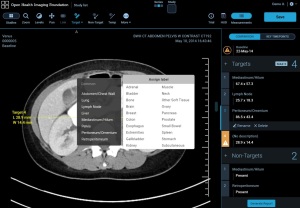
The OHIF Web Viewer is being used to help diagnose cases of COVID-19.
Users of a web-based imaging platform developed by Massachusetts General Hospital and its partners are shifting its use from identifying cancers to detecting and comparing medical images depicting COVID-19.
Initially designed for cancer imaging research and trials, the Open Health Imaging Foundation web viewer and its underlying Cornerstone libraries are being used for analysis in a series of projects aiming to provide faster diagnoses and care for COVID-19.
“This viewer provides performance that you typically only get from an installed [software] application, but we do it through a web browser,” said Dr. Gordon J. Harris, the corresponding author of an upcoming paper about the viewer, in a statement. "This is a free, open-source extendable platform that is already being used by projects worldwide.”


Ad Statistics
Times Displayed: 19306
Times Visited: 51 Stay up to date with the latest training to fix, troubleshoot, and maintain your critical care devices. GE HealthCare offers multiple training formats to empower teams and expand knowledge, saving you time and money.
Among the COVID-19 projects the OHIF Viewer and/or its underlying Cornerstone libraries are being used for are the DetectED-X CovED virtual clinical environment platform from Australia, which educates radiologists on CT depictions of COVID-19; the VUNO MedLung Quant and VUNO Med Chest X-ray AI programs in South Korea for diagnosing the virus; and the Nextcloud DICOM Viewer from Germany and Brazil, an open source, cloud-based and web-based medical image viewer for diagnosing COVID-19 from sites across Brazil fast and securely.
The interoperable and commercial grade program is a zero foot software that can be run in a web browser and spares users the hassle of downloading any software. It can be launched on a web server on a local computer or in the cloud, and is accessible from multiple locations. Researchers can freely download, modify, and contribute to the source code for the program, which has led to it being downloaded more than 8,500 times and translated into several languages.
The Precision Imaging Metrics Program developed by MGH and the Dana-Farber/Harvard Cancer Center has facilitated adoption of the user-friendly program by a number of NCI-designated cancer centers who rely on it to perform more than 25,000 oncology imaging assessments annually for over 1,000 active clinical trials. They include Dana-Farber/Harvard Cancer Center; Yale Cancer Center; Fred Hutchinson Cancer Research Center at University of Washington; Huntsman Cancer Institute at University of Utah; Winship Cancer Institute at Emory University; Massey Cancer Center at Virginia Commonwealth University; Medical College of Wisconsin; Karmanos Cancer Center at Wayne State University; and Nationwide Children’s Hospital.
“Many academic and industry projects are using the OHIF platform and associated Cornerstone tools for developing novel web-based imaging applications, and machine learning companies are now also tapping into it,” said Harris.
A number of companies are developing and deploying software programs to help providers not just diagnose COVID-19 but monitor its progression in patients. GE, for instance, recently
unveiled its Mural Virtual Care Solution to alert hospitals to when a patient’s condition is at risk of deterioration and identify those who require intervention, including ventilation and lung injury management for patients on extended mechanical ventilation support.
Harris’ paper on the viewer will be published in the
Journal of Clinical Oncology: Clinical Cancer Informatics.


