by
John R. Fischer, Senior Reporter | September 12, 2022
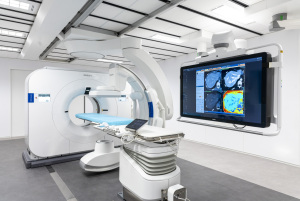
Philips has designed the world's first spectral detector angio CT suite.
Philips has combined its Spectral CT 7500 system and its Azurion image-guided therapy system with FlexArm to form the world’s first spectral detector angio CT suite.
Designed to perform whole-body imaging in 2D and 3D, Azurion with FlexArm utilizes imaging systems, software and specialized diagnostic and therapeutic solutions to visualize critical anatomy so that clinicians can perform complex procedures.
Spectral CT improves detection, delineation and quantification of lesions to better plan minimally invasive operations. It also has shown more sensitivity in detecting malignant findings and improving readings of incidental findings.


Ad Statistics
Times Displayed: 18179
Times Visited: 41 Stay up to date with the latest training to fix, troubleshoot, and maintain your critical care devices. GE HealthCare offers multiple training formats to empower teams and expand knowledge, saving you time and money.
Using Philips’ scanner allows clinicians to image patients only once to distinguish and quantify different tissues. Together, both solutions will allow clinicians to explore potential new treatments and improve minimally invasive procedures in oncology, stroke and trauma care.
"We cannot only visualize better but also quantify, for example, iodine uptake in the tumor with an embolization. Or using Spectral information to define treatment success before the patient leaves the room," Karim Boussebaa, general manager of image-guided therapy systems at Philips, told HCB News.
The company also announced new research collaborations around its combined systems. Leiden University Medical Center, in the Netherlands, for example, has joined its global network of clinical partners investigating possible treatments with both solutions.
"Adding spectral CT imaging to the interventional suite will enable us to offer new treatment opportunities, avoid moving patients from one imaging suite to another, and offer the unique benefits of spectral CT information when you need it,” said Dr. Mark Burgmans, head of interventional radiology at Leiden University Medical Center.
Philips is additionally working with the University of Pennsylvania to study the practicality of using spectral CT-based tissue temperature mapping for real-time feedback during tumor thermal ablation procedures. This will enable clinicians to confirm if the treatment was effective before the patient leaves the room, potentially reducing the risk of localized tumor recurrence.
A third institution is University of Insubria, in Italy, which has been studying the value of spectral CT for performing high-confidence tumor biopsies. The researchers involved in that project say the Spectral CT 7500 system can distinguish active from inactive regions in tumors, making it a better guide for clinicians during biopsies and reducing inconclusive results.
"For acute procedures such as stroke and traumatic bleedings we see a paradigm shift to a one-stop-shop approach, to reduce time between diagnosis and treatment. Physicians want to have diagnostic- and therapeutic capabilities in one room to minimize delays and improve outcomes," said Boussebaa.
The findings for that will be presented at the Cardiovascular and Interventional Radiological Society of Europe Annual Meeting in September 2022, in Barcelona.


