by
Brendon Nafziger, DOTmed News Associate Editor | November 03, 2009
Breast PET could allow
better detection of
secondary tumors
PET imaging is better at distinguishing benign from deadly lumps in the breast than MRI scans alone, according to a recent study sponsored by the National Institutes of Health.
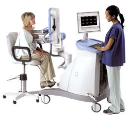
The three-year, multi-center trial found that positron emission mammography, or PEM, a new breast-specific PET modality, showed greater specificity than MR -- that is, it registered fewer false positives during diagnostic tests.
In the study, announced this week by Naviscan, the San Diego, Calif.-based developers of PEM, the researchers looked at almost 400 women recently diagnosed with breast cancer. The scientists wanted to see how PEM imaging, a modality that, according to Naviscan, only became commercially available a couple of years ago, fared against MR when searching for secondary or additional breast tumors, the nature of which can help doctors determine whether to remove just the cancerous tissue or the whole breast.


Ad Statistics
Times Displayed: 316
Times Visited: 2 Keep biomedical devices ready to go, so care teams can be ready to care for patients. GE HealthCare’s ReadySee™ helps overcome frustrations due to lack of network and device visibility, manual troubleshooting, and downtime.
PEM correctly tagged 151 of the 189 benign tumors as benign, an 80 percent success rate. On the same subjects, MRI was more likely to mistake benign tumors for malignant ones, with only a 66 percent success rate.
"Breast MRI has been very good where sensitivity has been concerned," Gary Seelhorst, a senior director of Naviscan, tells DOTmed News. But while MR can help doctors identify suspicious lesions, it's "notorious" for having quite low specificity for breast cancer, Seelhorst says.
Part of the reason for this lies in the way the technology works. In PEM, small doses of a radioactive pharmaceutical, fludeoxyglucose, are injected into the patient. This material fires out positrons that are detected by monitors the PEM places on either side of the tissue. But the gain in specificity comes in how fludeoxyglucose is processed by the body: generally, only malignant tumors will take it up, so it's quite easy for a physician to see on screen whether a lump is dangerous or not.
Increased sensitivity, too
The NIH study also found that the benefits of PEM weren't limited to specificity: by combining MR scans with PEM, sensitivity, or the ability to pick up any suspicious growths, rose by 20 percent. "There's an additive effect," says Seelhorst. "You pick up everything, you're going to know exactly where everything is."
And the reason for that is PEM can compensate for a known MR drawback: MRI scans can only really detect breast cancer that has vasculature, or has recruited blood vessels to feed itself. PEM has no such limitation.
Gentle learning curve, low price
PEM could have some more practical benefits, too. According to Seelhorst, his company's PEM machines, at around $600,000 each, undercut many MRI modalities by half a million to a million dollars.
Currently, Naviscan has around 40 PEM machines installed at centers, mostly in the U.S., but he hopes the number will grow, as his company is pushing for PEM to be taken on as, at least for now, a second-line diagnostic technology for women whose initial mammograms suggest a follow-up is needed.
"Radiologists, surgeons, oncologists are the ones starting to understand the importance of this newer technology," he says. "What we're trying to fight for right now is exposure."