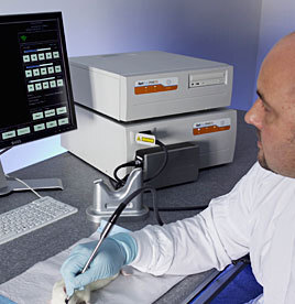
The Optiscan Five
pen microscope
A laser microscope that picks up fluorescently-dyed cells could result in more precise tumor extraction during brain surgery, and streamlined workflows in the operating room.
According to a study published recently in the Journal of Neurosurgery, neurosurgeons at the Barrow Neurological Institute at St. Joseph's Hospital and Medical Center in Phoenix, Ariz. found that the pen-sized microscope could help them precisely identify where tumor tissue ends and healthy tissue begins.
The microscope works by letting doctors detect the edges of cancerous tissue at the cellular level. The cancerous tissue is distinguished from healthy tissue by fluorescent dyes, which selectively tag the non-healthy growths.


Ad Statistics
Times Displayed: 656
Times Visited: 5 Fast-moving cardiac structures have a big impact on imaging. Fujifilm’s SCENARIA View premium performance CT brings solutions to address motion in Coronary CTA while delivering unique dose saving and workflow increasing benefits.
"There are a couple problems in the OR with respect to malignant brain tumors," Mark Preul, M.D., the director of the Neurosurgery Research Laboratory at Barrow Institute, tells DOTmed News. "One, the tumors are infiltratives so you don't exactly know their margins. You can image these things, but it doesn't give you a look at the actual cellular dimensions."
That's why in surgeries, while the patient lies with his head open, the surgeons will send samples of the tissue to the neuropathologist's lab so they can determine if the doctor managed to completely excise the lesion. But usually, this process takes about 40 minutes.
"The tissue has got to be mounted, stained, looked at; a report has to go back to the OR," says Dr. Preul. "There's a real workflow problem."
Plus, Dr. Preul says with frozen cutting-sections, you can get visual artifacts that can impede proper diagnosis, and can sometimes lead to mis-classification of tumor aggressiveness. As a surgeon, "you will tailor surgery according to pathological interpretations," he says, so it's important to know what sort of growth they're dealing with.
But with the device, known as a confocal microscope, working in conjunction with brain-safe dyes, Dr. Preul hopes doctors can accurately learn, on-the-fly, the extent of a tumor's growth, so they can easily, quickly and completely cut it out.
Finding brain-friendly fluorescent dyes
The laser microscope was developed by Optiscan, an Adelaide, Australia based company, which is collaborating with Carl Zeiss, a storied German optics firm out of Oberkochen, Germany.
Although it has been used in GI endoscopies for the past couple of years, it has only recently been applied to brain surgeries. One reason for the delay had been concern over fluorescent dyes.
"In the brain, there aren't many fluorescent markers that have been approved by the FDA for use," says Dr. Preul. The problem is that many fluorescent markers are suspected of damaging cellular DNA, says Dr. Preul. This isn't so much of a problem in the gut, where there's rapid tissue turnover around every two days, he says; but the brain works differently, so caution has to be used in anything introduced.
Although some safer dyes had been applied to the brain, they tended to also light up masses of surrounding blood cells, making distinguishing between tumor cells and blood cells difficult for the microscope, Dr. Preul says.
The answer appears to be a dye called 5-Aminolevulinic acid, or 5-ALA. "The 5 ALA really only lights up tumor cells," says Dr. Preul. "It's like having a topical fluorophor, except it doesn't light up erythrocytes [red blood cells]. All you see are the tumor cells."
Opening salvos
5-ALA is currently being tested in around six investigative centers in the U.S., and does not have FDA approval.
Also, the microscope technique is only in the early stages. According to Dr. Preul, only about 30 or 40 patients have had the procedure done on them.
"We're going to have years worth of work," says Dr. Preul, "where you acquire patients and then you gauge their surgical resection with the Optiscan versus surgical resection without it."
"We're only at the opening salvos," he adds.


