by
Brendon Nafziger, DOTmed News Associate Editor | December 21, 2010

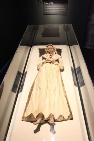
Veronica Orlovits
(PRNewsFoto/American
Exhibitions, Inc.,
Craig T. Mathew)
Radiology news from around the world for Dec. 21.
Technology could make processing virtual colonoscopies 10 times faster
Massachusetts General Hospital scientists say they can make computer processing of virtual colonscopies 10 times faster by relying on a mix of Hewlett-Packard PCs, Intel developer tools and Microsoft programs. Plus, the high-powered VC system could automatically edit out fecal matter in the colon, doing away with the obligatory night-before laxative cleanse.


Ad Statistics
Times Displayed: 656
Times Visited: 5 Fast-moving cardiac structures have a big impact on imaging. Fujifilm’s SCENARIA View premium performance CT brings solutions to address motion in Coronary CTA while delivering unique dose saving and workflow increasing benefits.
Traditional colonoscopies require patients under sedation to have a camera threaded through the colon, which has to be cleansed the night before. For virtual colonoscopies, CT scans create 3D images of the colon, but still a device has to be put in the rectum to inflate the colon with gas, and patients need to take laxatives so fecal matter doesn't obscure the reconstructed 3D image.
But Hiro Yoshida, who researches 3D imaging at Boston-based Massachusetts General's radiology department, says he developed a DTLS (datagram transport layer security) algorithm that cuts the time to processing the 3D images from half an hour to three minutes. It also can remove fecal matter digitally.
"Rather than take that chalky laxative and do the preparation the day before, what that does is automatically tag the items inside you and can flag the potential polyps -- it's done in a matter of minutes," said Steve Aylward, Microsoft's general manager for commercial health, in a conversation with eWEEK.
The VC-processing runs on HP multicore systems, Microsoft's Server 2008 and .NET 4.0 Framework, a Vectorform image viewer and Intel's Parallel Studio 2011, according to eWeek.
Yoshida said his team was still working out the kinks, but that the technology could be available for use as early as third quarter next year.
Hungarian mummy gets CT scan
Columbia St Mary's Hospital in Milwaukee had a patient scheduled for a CT scan on Dec. 10. The doctors had already diagnosed her with tuberculosis, but the exam wasn't urgent. The woman, Veronica Orlovits, was born more than 230 years ago.
Orlovits (born in 1770), her husband Michael and son Johannes were a trio of mummies on loan to the Milwaukee Public Museum from the Hungarian Natural History Museum in Budapest.
The family was one of a cache of mummies discovered in 1994 during renovation of a Dominican church in Vac, Hungary, just north of Budapest. Diggers found the remains in two burial crypts more than 300 years old, which were sealed in 1838. The crypt's cold, dry air and oil from the pine shavings lining the coffins helped preserve the bodies, according to the exhibitor Mummies of the World, which helped arrange for the mummies' visit.

