by
Brendon Nafziger, DOTmed News Associate Editor | July 17, 2012
Sodium buildup could serve as a biomarker for multiple sclerosis on MRI brain scans, according to a new French-led study.
The study, published online in the journal Radiology on Tuesday, found that total sodium concentrations increased in certain brain regions during early stages of the disease but spread over the entire brain in more advanced forms. Sodium accumulations, even in normal-seeming parts of the brain, also correlated with disease severity.
"Brain sodium MR imaging may help monitor the occurrence of tissue injury and disability," wrote the researchers. Wafaa Zaaraoui, with the National Center for Scientific Research in Marseilles, France, was the first author.


Ad Statistics
Times Displayed: 77597
Times Visited: 2716 Ampronix, a Top Master Distributor for Sony Medical, provides Sales, Service & Exchanges for Sony Surgical Displays, Printers, & More. Rely on Us for Expert Support Tailored to Your Needs. Email info@ampronix.com or Call 949-273-8000 for Premier Pricing.
Multiple sclerosis is an incurable disease thought to be caused by autoimmune attacks in the central nervous system on myelin, the fatty substance that covers nerve cells. The damage to myelin interferes with proper nerve signaling, leading to a host of motor, balance and cognitive problems.
Sodium MRI imaging is a type of MRI scan that draws data from the spin of nuclei in 23Na, rather than hydrogen nuclei. It's under investigation for applications from cartilage health to breast imaging and Alzheimer's disease.
How the study was done
In the study, the researchers examined 26 patients suffering from relapse-remitting multiple sclerosis, the most commonly diagnosed kind, which affects 85 percent of those diagnosed with MS, according to the National Multiple Sclerosis Society. This form of MS is characterized by occasional attacks, or relapses, followed by remissions.
Of these patients, 14 had early-stage disease, that is, the disease had lasted for less than 5 years, and 12 had advanced MS, which had lasted longer than 5 years. An additional 15 healthy controls were also included in the study.
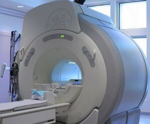
All participants received sodium MRI scans on a 3-Tesla machine. Total sodium concentrations were quantified for the brain's white matter and gray matter, as well as for MS lesions -- areas where myelin had been stripped. Disease severity was then measured by scores on the Expanded Disability Status Scale.
Results
According to the study, sodium concentrations increased in lesions stripped of myelin in all MS patients, but in patients with advanced disease, even normal-seeming gray and white matter showed higher levels of sodium buildup. Increased sodium concentrations were limited to the brainstem, cerebellum and temporal poles for early-stage patients, but were widespread for those who had the disease for more than 5 years, the study said.
Sodium buildup in gray matter and in the brain's motor networks also correlated with symptom severity, researchers said.

