by
Gus Iversen, Editor in Chief | November 19, 2014
From the October 2014 issue of HealthCare Business News magazine
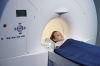
“You can measure that with MR and, through computer algorithms, find where the tracks run in the brain in x-y-z planes,” says Boop. DTI measures the restricted diffusion of water in tissue and can also be useful in collecting information about muscle.
Instead of dealing merely with white matter, DTI lets physicians distinguish one track from another. “Certain pathologies you can see if those tracks are pushed out of the way, spread, apart, disrupted,” says Boop, “and that can help with diagnosis in some situations.”


Ad Statistics
Times Displayed: 2147
Times Visited: 10 Fast-moving cardiac structures have a big impact on imaging. Fujifilm’s SCENARIA View premium performance CT brings solutions to address motion in Coronary CTA while delivering unique dose saving and workflow increasing benefits.
Functional MR, on the other hand, measures focal changes in blood flow in the cortex of the brain, explains Boop. When an area of the brain is active, the neurons increase their metabolism. The brain responds by increasing the blood flow, says Boop. If you flash a strobe light in front of someone’s eye and their visual cortex responds to that, you can measure those changes in blood flow over that area of the cortex and you know that’s the area that’s responsible for vision.
Knowing, for example, where that vision area exists in the brain helps the surgeon make sure to avoid it and not produce any unwanted side effects or cause additional damage during the procedure.
According to Dr. Arkadi Stolpner, president of the Diagnostic Treatment Center of the International Institute of Biological Systems, DTI is distinguished from fMRI in the physics used to obtain the data. “DTI is based on the speed and direction of water molecule movement while fMRI measures vascular response in neuronal activity,” says Stolpner.
Stolpner says fMRI is not a diagnostic tool but a way to identify activity in the cerebral cortex. At his treatment center they are currently conducting fMRI studies on three to six patients per week, but that number is always increasing. “The presence of fMRI stimulates the doctor’s desire to perform it for their patients,” says Stolpner, “It improves the final quality of surgery.”
Siemens is engaged with fMRI with their new MAGNETOM Prisma 3T, which was specially designed to manage the workload that comes with such concentrated imaging. “If you were doing a sleep study where the patient was asleep during the scan and you wanted to do it for an hour,” says Stuart Clarkson, the senior director of MR business with Siemens, “you’re actually putting a large current through these gradient coils and reversing it very quickly, and that generates a lot of heat.” Clarkson says the gradient coils on the Prisma feature an advanced cooling system that allows for a constant temperature and therefore helps to ensure accurate results.