by
Gus Iversen, Editor in Chief | November 19, 2014
From the October 2014 issue of HealthCare Business News magazine
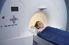
The Prisma also allows for a brand new MR technique called diffusion spectral imaging, (DSI). Clarkson says DSI is, “really the next level of trying to look at very subtle changes in crossing fibers of white matter. It allows us to do 514 different tensor directions.”
Where DTI is more of a structural study looking at white matter fibers and the way the cortex of the brain is attached to the spinal cord, DSI shifts that focus. “Sometimes when you have a voxel, there are many fibers that go through that one voxel,” says Clarkson. “To resolve them, to see the individual fibers, you would switch to DSI, which can differentiate the crossing fibers.” Voxels are pixels with volume; the points of data on an x-y-z plane.


Ad Statistics
Times Displayed: 135743
Times Visited: 7840 MIT labs, experts in Multi-Vendor component level repair of: MRI Coils, RF amplifiers, Gradient Amplifiers Contrast Media Injectors. System repairs, sub-assembly repairs, component level repairs, refurbish/calibrate. info@mitlabsusa.com/+1 (305) 470-8013
“You can think of DSI as an improvement on DTI,” says Vossough. “It’s helpful in pre-surgical mapping prior to removal of brain legions, brain tumors, and vascular malformations.” He says there have been many occasions when DSI revealed something to him that prompted him to contact the surgeon and re-route the trajectory of the planned operation.
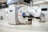
PET/MR – It has some believers, but still has a ways to go
The combination of PET and MR represents another exciting breakthrough in diagnostic image exploration. PET is often used to detect malignancies and, more and more, it is being used to diagnose Alzheimer’s disease. Historically, technologists have been able to take a PET and an MR and co-register the images after taking them individually; a technique which sometimes yields greater insight than either image on its own. Today that process is being done in
one fell swoop.
GE is seeking 510k approval for a hybrid system that would automatically put these data sets together so a patient would only need to take a single trip to the hospital. On behalf of the project, Hausmann says, “We are installing two systems in Europe as we speak, and three test systems for FDA approval.” The name of GE’s hybrid system is Signa PET/MR, which is a throwback to a previous line of GE scanners under the Signa product line.
For GE, software updates have always been a fundamental part of their product enhancements. It will be no different with PET/MR, which may be available as an upgrade to customers already using their high-end Discovery MR750w 3.0T scanner.
Clarkson says Siemens is currently the only company with an FDA approved PET/MR scanner; the Biograph mMR. Clarkson describes the benefits of PET/MR, “We’re looking at interesting studies where we can use MR data to correct PET data.” If a patient has Parkinson’s disease, for example, and their head is very hard to hold still, your PET images will not have satisfactory resolution. With Biograph mMR, Clarkson says, “You can acquire MR data very quickly and then actually register the movement of the patient’s head and correct the PET data to account for the motion.”