by
Christina Hwang, Contributing Reporter | April 18, 2016

Hope to better understand
human genes by altering the
corresponding gene in tadpole
Courtesy: Yale
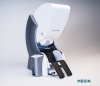
Scientists from Yale University have developed a multimodal imaging system that can look at both the structure of biological tissue and the internal activities of a body.
In one mode, the laser is able to reduce the amount of grainy, random speckle that can potentially distort the resulting image. Speckle is usually found in the light emitted by traditional lasers that conduct high-speed imaging.
“The speckle-free images are taken to reveal the structure of a biological object, similar to the images taken with the illumination of the lamp in a conventional microscope,” Hui Cao, a professor of applied physics and of physics at Yale and corresponding author of the study, told HCB News.


Ad Statistics
Times Displayed: 346960
Times Visited: 21062 MIT labs, experts in Multi-Vendor component level repair of: MRI Coils, RF amplifiers, Gradient Amplifiers Contrast Media Injectors. System repairs, sub-assembly repairs, component level repairs, refurbish/calibrate. info@mitlabsusa.com/+1 (305) 470-8013
Speckle can provide additional information since changes in its pattern can be used for biological imaging. “In the other mode, laser speckles are generated from the moving sample that allows one to probe the dynamic activity – such as a heartbeat or movement of a cell,” Cao said.
The initial testing was done on a living tadpole, and two co-authors of the paper, Michael Choma and Mustafa Khokha from Yale Medical School, worked for several years developing and using imaging to study congenital heart disease genes.
Cao said the study on tadpoles is important because new genetic tools are revealing that many human genes may be involved in congenital heart disease.
The laser has not been tested on other organisms but Choma will be using the laser to perform biopsies on the lungs of certain specimens, by studying cilia, the lining of the airway.
“We currently lack the lab and clinical diagnostic tools to precisely understand mucus flow physiology and the specific details of how it impacts different diseases,” Cao said, and the goal is to do high-performance imaging of these human biopsy specimens using the imaging systems that the researchers have developed.
Choma explained in a statement that the relationship between structure and function is fundamental to the study of biology and it is particularly true when studying micro-scale motions and flows within living tissue.