by
Lauren Dubinsky, Senior Reporter | October 26, 2016
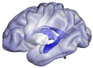
Computational model of the cortical
and subcortical brain structures that
form the basis of the BrainPrint
A team of researchers at Massachusetts General Hospital (MGH) have found a way to spot individuals in the early presymptomatic stages of Alzheimer’s disease with MR. The scans revealed that those with the disease had asymmetrical brain structures in the left and right sides of the brain.
“Compared to the size of a structure, the geometric shape can pick up subtle changes in sub-structures much earlier,” Martin Reuter, lead researcher, told HCB News. “Lateral asymmetries in dementia may have been overlooked for that reason in the past.”
In particular, the asymmetry of the hippocampus and amygdala increases as the disease becomes more severe. The researchers believe the asymmetry of those and a few other structures could be a biomarker for detecting early-stage dementia.


Ad Statistics
Times Displayed: 346433
Times Visited: 21062 MIT labs, experts in Multi-Vendor component level repair of: MRI Coils, RF amplifiers, Gradient Amplifiers Contrast Media Injectors. System repairs, sub-assembly repairs, component level repairs, refurbish/calibrate. info@mitlabsusa.com/+1 (305) 470-8013
The team developed a computer-aided system called BrainPrint that represents the whole brain based on the shapes, rather than the size or volume, of the structures. An article published in 2015 in the journal,
NeuroImage, showed that BrainPrint can be as accurate as a fingerprint in distinguishing individuals.
This new study used BrainPrint to evaluate structural asymmetries in MR images of 700 patients involved in the National Institute of Health-sponsored Alzheimer’s Disease Neuroimaging Initiative (ADNI). The participants received an MR exam at enrollment and then again every six to 12 months, along with cognitive and genetic testing.
The researchers assessed the data from ANDI participants that underwent at least three MR exams. They were then divided into four groups: those diagnosed with probable Alzheimer’s when entering the study, healthy controls with no sign of dementia, those with mild cognitive impairment (MCI) that remained stable over two to three years and those with MCI that progressed to Alzheimer’s.
BrainPrint revealed that differences in the shapes of the hippocampus and amygdala were highest in individuals with dementia and lowest in the healthy controls. Among the group that was diagnosed with MCI, the baseline asymmetry was higher in those that developed Alzheimer’s, and increased even more as symptoms became apparent.
“Clinical Alzheimer’s disease diagnosis occurs very late. By that time decades of advanced neurodegeneration have taken their toll and large portions of the brain are atrophied,” said Reuter. “Recovery at this stage is unlikely, even stopping or significantly slowing the neurodegenerative process at this time will be very difficult.”
If presymptomatic or early-stage individuals at risk of developing the disease are identified, they can be recruited into studies investigating novel therapies, because preventive treatment can still be successful.
Reuter and his team are planning to further explore the relationship between brain asymmetries and established Alzheimer’s disease biomarkers to get a better understanding of the underlying biological mechanisms.
“This approach is a building block for spotting early-stage Alzheimer’s,” he said. ”For high-accuracy Alzheimer’s prediction, genetic markers, other imaging markers, as well as test scores, lifestyle, nutrition, etc., will need to be considered jointly.”


