by
Lauren Dubinsky, Senior Reporter | January 17, 2017

Multiplexed image analysis with HySP
Courtesy of Francesco Cutrale
By taking a broader, more complete view of disease, researchers at the University of Southern California believe that their "5-D image analysis technique" called Hyper-Spectral Phasor analysis could usher in an new era in diagnostics — one where cell phone images could be the front line of care.
Fluorescent imaging is used to locate proteins and other molecules in cells and tissues by tagging the molecules with dyes that glow under certain light. It helps scientists find out which molecules are produced in large amounts in cancer or other diseases and that can be used to diagnose or identify therapeutic drugs.
However, only evaluating one or two molecules doesn’t give scientists a clear understanding of how those molecules behave in the real world. To get that view, scientists need to watch a target move over time.


Ad Statistics
Times Displayed: 346433
Times Visited: 21062 MIT labs, experts in Multi-Vendor component level repair of: MRI Coils, RF amplifiers, Gradient Amplifiers Contrast Media Injectors. System repairs, sub-assembly repairs, component level repairs, refurbish/calibrate. info@mitlabsusa.com/+1 (305) 470-8013
Historically, researchers had to look at different molecules separately then apply complicated techniques to layer them together, which was time-consuming and expensive. But with HySP, they can look at all of those molecules at once.
“Biological research is moving toward complex systems that extend across multiple dimensions, the interaction of multiple elements over time,” said postdoctoral fellow Francesco Cutrale, in a statement.
Cutrale developed HySP with Scott Fraser, Elizabeth Garrett chair in Convergent Bioscience and provost professor of Biological Science.
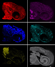
Different fluorescent light wavelengths reveal
features of a zebra fish embryo
Courtesy of Francesco Cutrale
Researchers at USC, Keck School of Medicine, Caltech and the University of Cambridge in the UK tested HySP on zebra fish and it worked extremely well. In the experimental model, they can use genetic manipulation to label the molecules, but that can’t be done in humans.
Natural fluorescence from biomolecules usually interferes with imaging, but a new computer algorithm can find weak signals in a cluttered background. The researchers can then find their targets within the body.
They plan to test the technique in the next couple of years on soldiers whose lungs were damaged by chemicals and irritants that they encountered in combat. A light-emitting probe will be extended down the soldiers’ lungs while the probe records images of the fluorescence in the surrounding tissue.
HySP will then be used to create a fluorescent map and it will be compared with healthy lung tissue. If the study is successful, they plan to further develop the technology so these soldiers and other lung patients receive more targeted treatment.
The researchers believe that HySP may also be used to analyze cell phone pictures of skin lesions to determine if they are at risk of being cancerous. It can look for any changes in the color or shape of the lesion over time.


Bill Gustafson
Enhanced Imaging
January 18, 2017 03:59
I think anytime you can expand or build on existing technology it is wise to investigate it.
to rate and post a comment