by
Thomas Dworetzky, Contributing Reporter | February 23, 2017
A new system has been developed that can do real-time MR image analysis aided by supercomputers.
This could cut patient callbacks, health care costs and boost precision medicine.
The automated platform can do in-depth MR scan analysis “in minutes,” according to a statement by researchers from the Texas Advanced Computing Center (TACC), the University of Texas Health Science Center (UTHSC) and Philips Healthcare


Ad Statistics
Times Displayed: 120730
Times Visited: 6941 MIT labs, experts in Multi-Vendor component level repair of: MRI Coils, RF amplifiers, Gradient Amplifiers Contrast Media Injectors. System repairs, sub-assembly repairs, component level repairs, refurbish/calibrate. info@mitlabsusa.com/+1 (305) 470-8013
The researchers showed their work in a proof-of-concept demonstration at the International Conference on Biomedical and Health Informatics recently in Orlando, Florida.
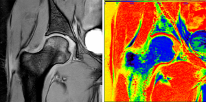
In their Florida demonstration UTHSC ran MR scans of a patient with a cartilage problem, to assess the state of the disease.
The project used a Philips MR scanner and the Stampede supercomputer. Communication between them was done using the TACC-developed Agave application program interface (API) platform infrastructure – basically a set of protocols and tools to exchange data and job control information. The scanner data was fed into a proxy server that ran a UTHSC-developed analysis tool, the GRAphical Pipelines Environment, or GRAPE.
The GRAPE analyzes scanned tissue and then sends information and suggestions back to the scanner operator.
For example, it might point out the need for a re-scan due to patient movement, or advise pursuing additional scans to check out a particular pathological issue.
The additional time resulting from this process was described as “minimal.”
"The Agave Platform brings the power of high-performance computing into the clinic," William Allen, a life science researcher for TACC and lead author on the paper, said in a statement. "This gives radiologists and other clinical staff the means to provide real-time quality control, precision medicine, and overall better care to the patient."
"We are very excited by this fruitful collaboration with TACC," said Refaat Gabr, an assistant professor of Diagnostic and Interventional Imaging at UTHSC and the lead researcher on the project. "By integrating the computational power of TACC, we plan to build a completely adaptive scan environment to study multiple sclerosis and other diseases."
The GRAPE interface has a “pipeline development toolbar” on the left of the window. On the right is a “canvas” on which the user can create a pipeline that controls the analysis of the scan.
This system could also permit other information to be extracted from an MR scan.
"There are a few thousand textures that can be quantified on MR [images]. These textures can be combined using appropriate mathematical models for radiomics. Combining radiomics with genetic profiles, referred to as radiogenomics, has the potential to predict outcomes in a number of diseases, including cancer, and is a cornerstone of precision medicine," according to Ponnada Narayana, Gabr's co-principal investigator and the director of Magnetic Resonance Research at the University of Texas Medical School at Houston.
This MR application is just one of the uses for "science as a service" platforms like Agave in medicine.
"Here we demonstrated this is possible for MR [images],” noted Allen. “But this same idea could be extended to virtually any medical device that gathers patient data," he observed, adding that, “in a world of big health data and an almost limitless capacity to compute, there is little reason not to leverage high-performance computing resources in the clinic."

