by
Lauren Dubinsky, Senior Reporter | July 18, 2017
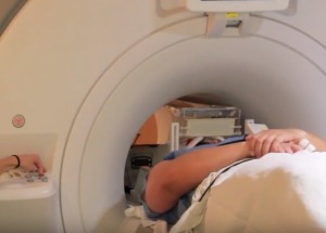
Two new studies published in The American Journal of Psychiatry uncovered that functional MR imaging can pave the way for personalized PTSD treatment.
“Individual patients differ with respect to these brain functions, hence identifying them, and identifying who is stronger in these capacities, can allow us to predict better who will and will not respond to psychotherapy in its current form,” Dr. Amit Etkin, senior author of the studies and associate professor at Stanford University Medical Center, told HCB News.
Not all PTSD patients benefit from treatment and about a third drop off it, according to Etkin, who is also an investigator at the Veterans Affairs Palo Alto Health Care System. Furthermore, about two-thirds receiving prolonged exposure therapy see a 50 percent reduction in symptoms, and 40 percent achieve remission.


Ad Statistics
Times Displayed: 7172
Times Visited: 104 Fast-moving cardiac structures have a big impact on imaging. Fujifilm’s SCENARIA View premium performance CT brings solutions to address motion in Coronary CTA while delivering unique dose saving and workflow increasing benefits.
To better understand how prolonged exposure therapy affects the brain, Etkin and his team used fMR to measure the brain activity of 66 PTSD patients as they completed five tasks that tested various emotional and cognitive functions.
They were shown images of happy, neutral or sad faces or scenes depicting an event intended to induce negative emotions like an argument or physical violence. They have to either respond to questions about the images or try to control their response to the image’s content.
After the initial brain imaging, about half of the patients underwent nine to 12 prolonged exposure therapy sessions and the remainder did not. After that, all patients underwent the same emotional response and regulation tests while the research team measured brain activity.
The first study focused on whether brain activity levels before treatment could help predict which patients would respond well to prolonged exposure therapy. The researchers measured the activity of certain brain regions during the five tasks.
They found that patients with lower activity in the amygdala and higher activity in the frontal lobe while viewing fearful faces had less PTSD symptoms after therapy. Patients with greater activation in a deep region of the frontal lobe when ignoring conflicting emotion information also responded better to therapy.
“In this study we were able to [predict treatment response] with 95 percent accuracy,” said Etkin. “While obviously there are caveats with any study, and that high of a number should always be taken with caution, it is quite promising.”
The second study found that prolonged exposure therapy improved PTSD symptoms. About four weeks after therapy ended, fMR imaging showed higher activity in the frontopolar cortex, which was surprising since the amygdala is usually associated with emotional processes in PTSD.

